Nucleotide Excision Repair in Tumor Response to DNA-Damaging Anticancer Therapies: Molecular Pathology, Pharmacological Mechanisms, and Clinical Resistance
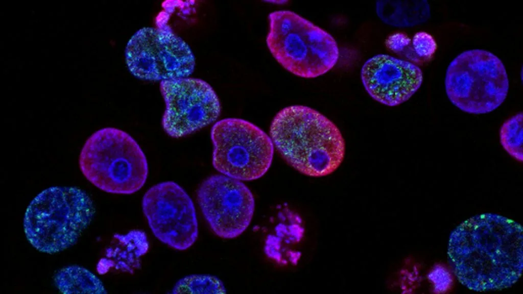
Nucleotide excision repair orchestrates the recognition and elimination of bulky DNA helix-distorting adducts induced by platinum-based chemotherapies and UV radiation. Dysregulation of this critical DNA damage response network directly dictates chemoresistance and therapeutic efficacy across multiple solid malignancies.
- Primary functional role: Dual incision of damaged DNA strands surrounding bulky lesions induced by platinum coordination complexes.
- Major clinical implication: Elevated ERCC1-XPF expression correlates with high platinum resistance and inferior progression-free survival.
- Targeted modulation: Inhibition of upstream damage recognition proteins (XPC, DDB2) or downstream repair endonucleases sensitizes refractory tumors to DNA-damaging agents.
Genomic instability driven by defective DNA repair mechanisms is a hallmark of human epithelial malignancies. The nucleotide excision repair (NER) pathway serves as the primary cellular defense mechanism against bulky genomic adducts.
Chemotherapeutic regimens relying on DNA interstrand and intrastrand crosslinks depend heavily on the functional status of the NER machinery for cytotoxicity. Tumors exploit NER upregulation to evade apoptosis induced by genotoxic stress.
CLINICAL & LABORATORY DIAGNOSTIC PROTOCOL
- Specimen acquisition: Formalin-fixed paraffin-embedded (FFPE) tumor tissue core biopsies or surgical resections fixed in 10% neutral-buffered formalin for 24 to 48 hours.
- Tissue processing: Microtome sectioning of FFPE blocks at 4-micron thickness onto positively charged adhesive glass slides, dried at 60 degrees Celsius for 60 minutes.
- Control standards: Certified positive control tissue with known high nuclear expression of ERCC1 and matching negative isotype control slides run concurrently.
Deparaffinization and Heat-Induced Epitope Retrieval (HIER)
Deparaffinize tissue sections through three changes of xylene for 5 minutes each, followed by rehydration through graded ethanol series (100%, 95%, 70%) to distilled water. Perform heat-induced epitope retrieval in a PT Link module using pH 9.0 Tris-EDTA buffer at 97 degrees Celsius for 20 minutes.
Peroxidase Blocking and Primary Antibody Incubation
Block endogenous peroxidase activity by incubating slides in 3 percent hydrogen peroxide solution for 10 minutes at room temperature, followed by a wash in Tris-buffered saline with 0.05 percent Tween-20 (TBST). Apply primary monoclonal antibody targeting ERCC1 (clone 8F1) at a 1:100 dilution and incubate for 30 minutes in a humidified chamber.
Polymer Detection and Chromogenic Development
Rinse slides thoroughly in TBST wash buffer and apply horseradish peroxidase (HRP)-conjugated secondary polymer detection reagent for 30 minutes at room temperature. Develop the chromogenic signal using 3,3-diaminobenzidine (DAB) substrate solution for 5 minutes, producing a stable brown nuclear precipitate.
Counterstaining, Dehydration, and Scoring
Counterstain nuclei with Mayer hematoxylin for 1 minute, blue in running tap water, dehydrate through graded ethanols, clear in xylene, and apply permanent mounting media with a glass coverslip. Score nuclear staining intensity and percentage of positive tumor cells using a validated semi-quantitative scale.
1. Pathophysiology, Etiology & Cellular Carcinogenesis
The nucleotide excision repair pathway operates through two distinct subpathways: global genome nucleotide excision repair (GG-NER) and transcription-coupled nucleotide excision repair (TC-NER). GG-NER is initiated by the **XPC**-RAD23B complex or **DDB2**-DDB1 heterodimer recognizing helix-distorting DNA lesions anywhere in the genome. TC-NER is triggered when RNA polymerase II stalls at a DNA lesion on the transcribed strand, recruiting **CSB** and **CSA** proteins.
Both subpathways converge at the recruitment of transcription factor IIH (**TFIIH**), containing DNA helicases **XPB** and **XPD**, which unwinds the DNA duplex around the damage site. Dual incisions are subsequently executed by the structure-specific endonucleases **ERCC1**-**XPF** 5 prime to the lesion and **XPG** 3 prime, releasing a 24-30 nucleotide oligomer containing the damage. DNA polymerase delta or epsilon, alongside DNA ligase I or III, fills and seals the resulting single-stranded gap.
Genomic instability arises when NER components are epigenetically silenced or mutated, leading to persistent mutagenic lesions that drive malignant transformation. Conversely, overexpression of NER genes in established tumors prevents apoptosis during platinum-based chemotherapy, fostering treatment resistance and disease progression across epithelial cancers.
2. Oncogenic Signal Transduction Pathways & Genomic Alterations
Upregulation of DNA repair genes is frequently linked to oncogenic signaling axes such as **PI3K**-**AKT**-**mTOR** and **RAS**-**RAF**-**MEK**-**ERK**. Hyperactivation of these pathways induces transcription factors that upregulate **ERCC1** and **XPF** mRNA expression, conferring enhanced tolerance to genotoxic stress. This coordinated stress response allows tumor cells to withstand replication fork stalling induced by DNA-damaging drugs.
Cell cycle checkpoint kinases **ATM** and **ATR** phosphorylate downstream targets that regulate NER incision kinetics during replication stress. Abrogation of these checkpoints via targeted small-molecule inhibitors forces cancer cells into premature mitosis with unresolved DNA adducts, resulting in mitotic catastrophe. Therapeutic synergy is achieved by combining DNA-damaging agents with **ATR** inhibitors such as berzosertib.
Secondary alterations within DNA damage response genes often emerge under selective pressure from alkylating agents and platinum compounds. Loss-of-function mutations in mismatch repair or homologous recombination genes frequently prompt compensatory upregulation of NER pathways, establishing a complex network of multi-drug resistance phenotypes in recurrent tumors.
3. Histopathological Grading, Morphological Subtypes & Differential Diagnosis
Malignancies subjected to platinum-based neoadjuvant therapy often exhibit distinct morphological alterations, including therapy-induced tumor regression, bizarre multinucleated giant cells, and extensive stromal hyalinization. Histopathological grading must account for residual viable tumor cellularity, quantified via the pCR (pathological complete response) or residual cancer burden (RCB) grading systems. High nuclear expression of **ERCC1** in pretreatment core biopsies frequently predicts poor histopathological response to platinum-based regimens.
Differential diagnosis of poorly differentiated carcinomas requires an extensive immunohistochemical panel to exclude non-epithelial mimics and define site-specific lineages. Diagnostic markers such as **TTF-1** and **Napsin A** identify pulmonary adenocarcinoma, whereas **p40** and **CK5/6** confirm squamous cell carcinoma lineages. Assessing **ERCC1** status within these diagnostic panels provides critical prognostic data regarding expected chemotherapy benefit.
Accurate pathological classification necessitates distinguishing primary therapy-resistant clones from therapy-naive architectures displaying intrinsic DNA repair proficiencies. Immunohistochemical evaluation of **XPF** and **ERCC4** expression levels helps pathologists stratify tumors into molecular subgroups sensitive to alternative non-platinum therapeutic strategies.
4. Targeted Therapeutics, Landmark Phase III Trials & Resistance Mechanics
Platinum-based coordination complexes, including **cisplatin** and **carboplatin**, form bulky 1,2-intrastrand d(GpG) crosslinks that physically distort the DNA double helix. These structural lesions are the primary substrate for nucleotide excision repair excision and removal. Overexpression of the **ERCC1**-**XPF** endonuclease complex directly correlates with rapid repair of these crosslinks and clinical resistance.
Landmark Phase III clinical trials, such as the **ICOG** and **E1505** trials in non-small cell lung cancer, demonstrated that **ERCC1** expression levels dictate overall survival benefit from platinum doublet chemotherapy. Patients with low **ERCC1** expression derived significant survival advantages from cisplatin-based adjuvant therapy, whereas high **ERCC1** expressors showed no benefit. Similar predictive correlations have been established in advanced ovarian and colorectal carcinoma trials.
Tertiary resistance to DNA-damaging therapies involves compensatory upregulation of translesion synthesis polymerases, such as **POLH**, which bypass remaining unresolved lesions. Novel therapeutic strategies incorporate **ERCC1**-**XPF** protein-protein interaction inhibitors and **PARP** inhibitors to abrogate compensatory repair pathways and re-sensitize refractory tumors to cytotoxic chemotherapy.
5. Molecular Staging, Minimal Residual Disease (ctDNA) & Prognostic Stratification
Integration of the **AJCC 8th Edition** TNM staging parameters with molecular biomarker profiling provides precise prognostic stratification for patients receiving genotoxic chemotherapy. Quantitative assessment of DNA repair transcripts alongside traditional staging metrics refines the prediction of recurrence risk in early-stage solid tumors. High expression scores of NER-associated genes correlate with advanced nodal involvement and reduced disease-free survival.
Liquid biopsy surveillance utilizing circulating tumor DNA (**ctDNA**) allows real-time tracking of minimal residual disease and clonal evolution during therapy. Detection of persistent mutant **ctDNA** allele frequencies following platinum-based chemotherapy signifies active subclonal resistance driven by enhanced DNA repair capacity. Serial **ctDNA** assays enable clinicians to detect treatment failure months before radiographic progression.
Prospective clinical paradigms increasingly utilize molecular signatures of DNA repair proficiency to guide personalized treatment escalations or de-escalations. Stratifying patients by comprehensive genomic profiling ensures that tumors proficient in nucleotide excision repair are directed toward non-platinum targeted therapies or immunotherapy combinations.