Oncological Emergencies in Cellular Pathology & Histology: Molecular Mechanisms, Histopathologic Diagnosis, and Acute Interventions
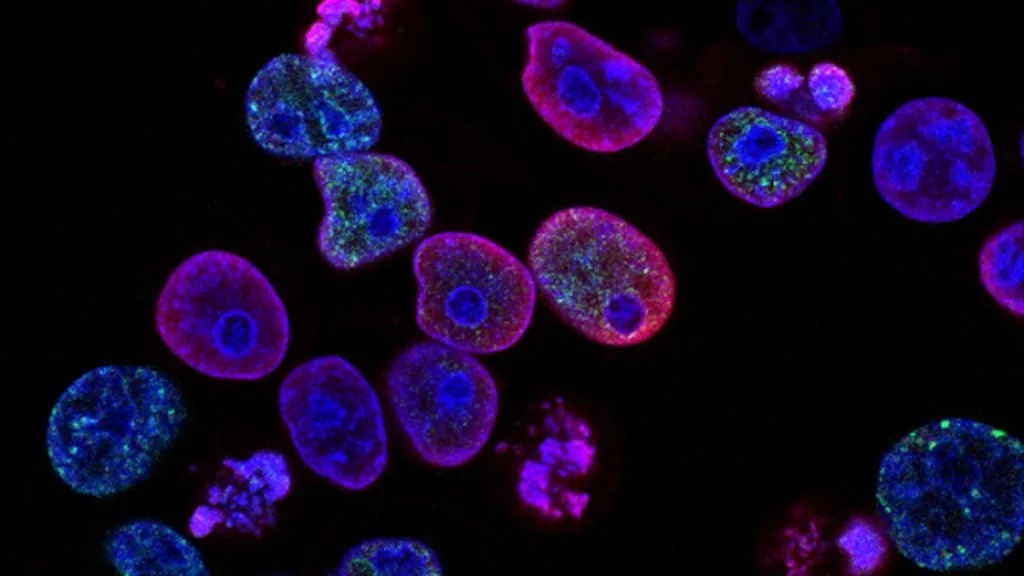
Oncological emergencies represent acute, life-threatening metabolic, mechanical, or structural complications arising directly from malignancy or cytotoxic therapy. Immediate cellular pathology and clinical pathology coordination are mandatory to establish rapid definitive diagnoses and prevent irreversible multi-organ failure.
- Primary pathology objective: Rapid histopathologic classification and molecular profiling of emergent tumor masses causing acute organ obstruction or compression.
- Metabolic emergencies: Tumor lysis syndrome and malignant hypercalcemia driven by cell turnover, **PTHrP** secretion, and acute tubular necrosis.
- Mechanical emergencies: Superior vena cava syndrome and spinal cord compression requiring urgent decompression and targeted systemic therapy.
Oncological emergencies occur in up to ten percent of advanced cancer populations, requiring rapid multidisciplinary intervention within cellular pathology and critical care units. Prompt tissue diagnosis differentiates primary hematologic malignancies from metastatic solid tumors presenting with acute obstruction or metabolic collapse.
Cellular pathology workflows must balance fixation fidelity with rapid turnaround times to identify actionable molecular targets under acute clinical distress. Delayed diagnosis directly correlates with permanent neurological deficits, renal failure, and early mortality in untreated patients.
CLINICAL & LABORATORY DIAGNOSTIC PROTOCOL
- Specimen acquisition: Urgent core needle biopsy or surgical decompression specimen fixed in 10% neutral-buffered formalin (NBF) within 15 minutes of devascularization.
- Tissue processing: Rapid microwave-assisted or standard automated tissue processing calibrated to maintain nuclear morphology and antigen preservation.
- Control standards: On-slide external positive control tissue and validated negative isotype control run in parallel with every emergency immunohistochemical batch.
Rapid Decalcification and Heat-Induced Epitope Retrieval (HIER)
Bake urgent sections at 60°C for 30 minutes, deparaffinize through sequential xylene baths, and rehydrate through graded ethanols. Perform HIER in target retrieval solution (pH 9.0 EDTA buffer) at 97°C for 20 minutes in a calibrated pressure module.
Endogenous Peroxidase Block & Primary Antibody Incubation
Quench endogenous peroxidase activity using 3% hydrogen peroxide for 5 minutes. Apply optimized primary monoclonal antibody (e.g., clone **SP263** for PD-L1 or clone **8D3** for TTF-1) at calibrated clinical dilution and incubate for 30 minutes at room temperature.
Polymer Detection System & DAB Chromogen Visualization
Rinse in Tris-buffered saline with 0.05% Tween-20 (TBST). Apply horseradish peroxidase (HRP)-conjugated anti-rabbit/mouse polymer secondary reagent for 20 minutes. Incubate with freshly prepared 3,3-diaminobenzidine (DAB) substrate-chromogen solution for 5 minutes until brown precipitates develop.
Counterstaining, Dehydration & Quality Control Scoring
Counterstain nuclei with Mayer hematoxylin for 1 minute, blue in running tap water, dehydrate through graded ethanols, clear in xylene, and coverslip with permanent non-aqueous mounting medium. Score tumor cell membrane and nuclear staining under light microscopy using validated scoring algorithm.
1. Pathophysiology, Etiology & Cellular Carcinogenesis
Rapid cellular proliferation and high mitotic indices predispose hematologic malignancies and germ cell tumors to spontaneous tumor lysis syndrome. Massive nucleic acid and electrolyte release overwhelms renal clearance capacity, precipitating acute kidney injury.
Epigenetic dysregulation and promoter hypermethylation silence regulatory tumor suppressor genes, driving unchecked cellular division and aggressive tissue invasion. Hypoxic microenvironments stimulate neoangiogenesis via vascular endothelial growth factor upregulation, leading to fragile, leaky vessels prone to hemorrhage.
Immunosuppressive stroma rich in regulatory T cells and myeloid-derived suppressor cells protects tumor beds from immune clearance. Cellular pathology analysis frequently reveals extensive geographic necrosis alongside actively proliferating invasive borders.
2. Oncogenic Signal Transduction Pathways & Genomic Alterations
Upstream receptor dimerization and autophosphorylation trigger sustained downstream signaling cascades through **RAS-RAF-MEK-ERK** and **PI3K-AKT-mTOR** pathways. These constitutive signals promote unchecked cellular survival, evasion of apoptosis, and increased metabolic demand in emergency presentations.
Genomic instability generates secondary driver mutations under therapeutic pressure, driving treatment failure and rapid clinical deterioration. Alterations in homologous recombination repair genes accelerate mutational burden and structural genomic rearrangements.
Bypass signaling pathways activated through parallel receptor tyrosine kinases maintain cellular viability despite targeted inhibition. This molecular adaptation underpins acute clinical resistance and rapid tumor progression.
3. Histopathological Grading, Morphological Subtypes & Differential Diagnosis
Architectural growth patterns including solid, micropapillary, and diffuse sheets characterize poorly differentiated malignancies presenting as oncological emergencies. High-power examination reveals marked nuclear pleomorphism, atypical mitotic figures, and extensive tumor necrosis.
Diagnostic immunohistochemical panels utilizing **TTF-1**, **Napsin A**, and **CK7** differentiate pulmonary adenocarcinomas from squamous cell carcinoma and lymphoma. Neuroendocrine markers such as **synaptophysin** and **chromogranin A** identify aggressive small cell carcinomas.
Differential diagnosis requires separating true oncological emergencies from acute infectious processes and autoimmune vasculitis. Accurate histologic subtyping dictates immediate therapeutic selection and dictates prognosis.
4. Targeted Therapeutics, Landmark Phase III Trials & Resistance Mechanics
First-, second-, and third-generation targeted kinase inhibitors provide rapid cytoreduction in molecularly selected patient populations presenting with impending organ failure. Therapeutic agents such as **osimertinib** and **crizotinib** achieve rapid objective response rates within days of administration.
Landmark Phase III clinical trials including **FLAURA** and **ADAURA** demonstrate profound progression-free survival benefits with third-generation inhibitors. Overall survival advantages are maintained across diverse patient subgroups harboring sensitizing mutations.
Tertiary resistance mutations such as **C797S** emerge following targeted therapy, necessitating fourth-generation inhibitors or antibody-drug conjugates. Combination strategies combining chemotherapy with immune checkpoint inhibitors address secondary resistance.
5. Molecular Staging, Minimal Residual Disease (ctDNA) & Prognostic Stratification
Integration of **AJCC 8th edition** TNM staging parameters with precise molecular biomarkers guides clinical decision-making during acute admissions. High tumor mutational burden and specific genomic alterations define high-risk cohorts requiring intensive inpatient monitoring.
Liquid biopsy surveillance utilizing circulating tumor data and cell-free DNA tracks minimal residual disease dynamics in real time. Serial monitoring detects subclinical therapeutic resistance before radiographic disease progression occurs.
Overall survival prognostic stratification combines clinicopathologic parameters with genomic profiles to guide post-emergency consolidation therapy. Prospective clinical trial paradigms continue to refine risk-adapted management strategies for acute oncological presentations.