Anaplasia in Cellular Pathology: Morphological Hallmarks, Genetic Instability & Oncogenic Reversion
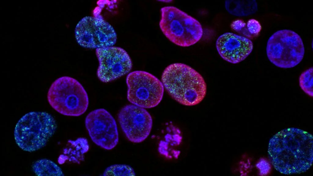
Anaplasia represents the hallmark architectural and cytological dedifferentiation of malignant neoplasms, characterized by severe cellular pleomorphism, hyperchromatic nuclei, and loss of functional tissue organization. This cellular phenotype correlates directly with high-grade malignancy, aggressive clinical trajectories, and intrinsic therapeutic resistance across solid tumors.
- Primary cytological feature: Marked cellular pleomorphism, bizarre multi-lobed nuclei, and atypical mitotic figures.
- Genomic underpinning: Complex chromosomal instability, frequent TP53 mutations, and large-scale copy number alterations.
- Clinical implication: Direct correlation with advanced AJCC staging, high mitotic rate (Ki-67 >= 50%), and aggressive recurrence.
Anaplasia occurs across diverse organ systems, predominantly affecting older adults with significant cumulative tobacco or carcinogen exposure. The histological manifestation indicates a profound regression from mature, specialized parenchymal architecture to a primitive, embryonic-like cellular state.
The underlying biological driver involves catastrophic genomic instability driven by defective DNA damage response pathways and unchecked cell cycle progression. This structural collapse abolishes normal tissue homeostasis and fuels rapid clonal evolution within the advancing tumor front.
CLINICAL & LABORATORY DIAGNOSTIC PROTOCOL
- Specimen acquisition: Incisional or excisional surgical biopsy fixed immediately in 10% neutral-buffered formalin (NBF) with a cold ischemia time strictly under 60 minutes.
- Tissue processing: Automated dehydration, clearing, and paraffin embedding with 3 to 4-micron microtome sectioning mounted on positively charged silanized glass slides.
- Control standards: Internal normal stromal elements serving as positive internal control alongside standardized external cell block controls for immunohistochemical validation.
Deparaffinization and Heat-Induced Epitope Retrieval (HIER)
Bake slides at 65°C for 45 minutes, pass through xylene (3 changes, 3 minutes each), and rehydrate through graded ethanols (100%, 95%, 70%) to distilled water. Perform HIER using a high-pH Tris-EDTA buffer (pH 9.0) in a pressure cooker maintained at 110°C for 15 minutes.
Endogenous Enzyme Block and Blocking Buffer Incubation
Block endogenous peroxidase activity by immersing slides in 3% aqueous hydrogen peroxide for 10 minutes at room temperature. Rinse thoroughly in Tris-buffered saline with 0.025% Triton X-100 (TBST) and apply protein blocking reagent for 10 minutes.
Primary Antibody Application and Amplification
Apply validated prediluted monoclonal antibodies against cell cycle and differentiation markers (e.g., Ki-67 clone MIB-1, p53 clone DO-7) and incubate in a humidified chamber at 25°C for 60 minutes. Apply secondary horseradish peroxidase-labeled polymer detection reagent for 30 minutes.
Chromogenic Development, Counterstaining, and Evaluation
Incubate sections with freshly prepared 3,3-diaminobenzidine (DAB) substrate for 5 minutes to visualize positive protein expression as a brown precipitate. Counterstain with Mayer hematoxylin for 45 seconds, dehydrate, clear in xylene, and coverslip using synthetic resin.
1. Pathophysiology, Etiology & Cellular Carcinogenesis
Anaplasia develops through progressive mutational accumulation that dismantles cellular differentiation programs and locks the cell in a perpetual proliferative state. The cell-of-origin undergoes severe transcriptional reprogramming, downregulating tissue-specific genes while upregulating embryonic stem cell markers.
Epigenetic dysregulation, characterized by global DNA hypomethylation coupled with targeted promoter hypermethylation of tumor suppressor genes, accelerates this dedifferentiation process. Checkpoint failures at the G1/S and G2/M transitions allow cells with damaged DNA to replicate continuously without undergoing apoptosis.
The surrounding tumor microenvironment adapts to this aggressive phenotype through extensive desmoplasia, hypoxia-induced factor 1-alpha (HIF-1alpha) signaling, and recruitment of immunosuppressive regulatory T cells. This altered stroma provides biochemical and mechanical cues that sustain the rapid expansion of anaplastic cell clones.
2. Oncogenic Signal Transduction Pathways & Genomic Alterations
Genomic profiling of anaplastic tumors frequently reveals devastating structural variants, including chromothripsis, whole-genome doubling, and complex translocations. These genomic catastrophes deregulate primary tyrosine kinase receptors and downstream second messenger cascades.
Constitutive hyperactivation of the **RAS-RAF-MEK-ERK** and **PI3K-AKT-mTOR** pathways drives unchecked cellular proliferation and inhibits pro-apoptotic signaling networks. Concurrently, biallelic inactivation of **TP53** and **RB1** removes critical genomic integrity checkpoints.
Secondary feedback loops and bypass tracks render anaplastic clones notoriously resistant to standard targeted therapies and cytotoxic agents. The loss of functional cell adhesion molecules further facilitates tissue invasion and metastatic dissemination.
3. Histopathological Grading, Morphological Subtypes & Differential Diagnosis
Histopathological examination reveals extreme cellular and nuclear pleomorphism, featuring bizarre, giant, multi-nucleated tumor cells alongside spindle-shaped and round undifferentiated variants. Mitotic figures are abundant and frequently atypical, displaying tripolar or multipolar spindles.
Immunohistochemical panels are essential to confirm epithelial, mesenchymal, or neuroendocrine lineage when morphology is entirely undifferentiated. Diagnostic markers such as **cytokeratins (AE1/AE3)**, **vimentin**, and lineage-specific transcription factors differentiate true anaplasia from lymphoma, melanoma, or sarcoma.
The differential diagnosis requires excluding reactive atypia, radiation-induced cellular changes, and severe dysplasia, which lack the true invasive architecture and clonal genetic aberrations definitive of anaplastic malignancy.
4. Targeted Therapeutics, Landmark Phase III Trials & Resistance Mechanics
Therapeutic management of anaplastic tumors relies on aggressive multi-modality regimens combining high-dose cytotoxic chemotherapy and advanced radiotherapy. Platinum agents and taxanes remain foundational, though objective response rates are frequently limited by primary resistance.
Landmark clinical trials, such as those evaluating immune checkpoint inhibitors like **pembrolizumab** and **nivolumab**, demonstrate durable responses in a subset of tumors exhibiting high tumor mutational burden or mismatch repair deficiency. Precision oncology approaches increasingly utilize comprehensive next-generation sequencing to identify actionable fusion proteins or rare targetable mutations.
Acquired resistance manifests rapidly via clonal selection of preexisting drug-tolerant persister cells or secondary resistance mutations that bypass targeted pathway inhibition. Combination therapies addressing multiple signaling nodes simultaneously represent the current frontier in overcoming this refractoriness.
5. Molecular Staging, Minimal Residual Disease (ctDNA) & Prognostic Stratification
Integrating **AJCC 8th Edition** TNM staging with high-grade histological anaplasia provides essential prognostic stratification for clinical decision-making. High tumor grade directly elevates risk categories across virtually all organ-specific staging systems.
Liquid biopsy surveillance utilizing circulating tumor DNA (**ctDNA**) offers a non-invasive modality to monitor minimal residual disease and detect early clonal evolution during or after therapy. Serial quantification of mutant allele frequencies correlates strongly with impending clinical recurrence.
Overall survival prognostic stratification remains poor for classical anaplastic variants, necessitating ongoing enrollment in early-phase clinical trials investigating novel cell-cycle inhibitors and bispecific immunotherapies.