Aurora Kinase A (AURKA) in Cellular Pathology: Amplification Mechanics, Cell Cycle Checkpoint Failure, and Targeted Alisertib Inhibitor Resistance
Aurora kinase A (**AURKA**) is an essential serine/threonine kinase mapped to chromosome 20q13.2 that governs mitotic entry, centrosome maturation, and bipolar spindle assembly. Overexpression or genetic amplification of **AURKA** drives chromosomal instability, aneuploidy, and oncogenic transformation across aggressive human carcinomas.
- Primary molecular driver: Chromosomal amplification at locus 20q13.2 resulting in constitutive **AURKA** protein overexpression.
- Targeted pharmacotherapy: Selective **AURKA** inhibitors such as **alisertib** disrupting spindle assembly checkpoint dynamics.
- Primary mechanism of resistance: Upregulation of compensatory mitotic kinases (**AURKB**, **PLK1**) and multidrug resistance efflux pumps.
Upregulation of **AURKA** is frequently documented in high-grade breast carcinomas, small cell lung cancer, and castration-resistant prostate cancer. Deregulation of this mitotic kinase directly correlates with advanced clinical stage, high mitotic index, and poor overall survival.
Cellular transformation driven by **AURKA** hyperactivation bypasses standard G2/M cell cycle arrest mechanisms. Unchecked kinase activity promotes abnormal centrosome amplification and unequal chromosomal segregation during mitosis.
CLINICAL & LABORATORY DIAGNOSTIC PROTOCOL
- Specimen acquisition: Core needle biopsy or surgical resection fixed in 10% neutral-buffered formalin (NBF) within 30 minutes of devascularization (cold ischemia time < 1 hr).
- Tissue processing: Formalin fixation time strictly calibrated to 24-48 hours at room temperature; paraffin embedding with 4-micron microtome sectioning on positively charged slides.
- Control standards: On-slide external positive control tissue (tonsil or high-grade placenta) and validated negative isotype control run in parallel with every analytical batch.
Tissue Deparaffinization & Heat-Induced Epitope Retrieval (HIER)
Bake slides at 60°C for 60 minutes, deparaffinize through sequential xylene baths (3 x 5 min), and rehydrate through graded ethanols (100%, 95%, 70%) to deionized water. Perform HIER in target retrieval solution (pH 9.0 EDTA buffer) at 97°C for 20 minutes in a calibrated PT Link module.
Endogenous Peroxidase Block & Primary Antibody Incubation
Quench endogenous peroxidase activity using 3% hydrogen peroxide for 10 minutes. Apply optimized primary monoclonal antibody targeting **AURKA** (e.g., clone 4C4) at calibrated clinical dilution of 1:100 and incubate for 60 minutes at room temperature in a humidified chamber.
Polymer Detection System & DAB Chromogen Visualization
Rinse in Tris-buffered saline with 0.05% Tween-20 (TBST). Apply horseradish peroxidase (HRP)-conjugated anti-rabbit/mouse polymer secondary reagent for 30 minutes. Incubate with freshly prepared 3,3-diaminobenzidine (DAB) substrate-chromogen solution for 5-10 minutes until brown precipitates develop.
Counterstaining, Dehydration & Quality Control Scoring
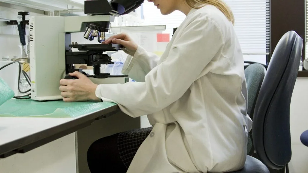
Counterstain nuclei with Mayer hematoxylin for 1-2 minutes, blue in running tap water or 0.08% ammonia water, dehydrate through graded ethanols, clear in xylene, and coverslip with permanent non-aqueous mounting medium. Score tumor cell nuclear and pericentrosomal staining under light microscopy using validated scoring algorithm.
1. Pathophysiology, Etiology & Cellular Carcinogenesis
Cellular transformation initiated by **AURKA** dysregulation stems from aberrant centrosome duplication cycles. Ectopic kinase activity triggers premature separation of centrioles during S-phase, generating multipolar mitotic spindles.
Epigenetic dysregulation and promoter demethylation at the 20q13.2 locus frequently drive transcriptional upregulation. This molecular cascade suppresses **TP53**-dependent apoptotic pathways, allowing genomically unstable cells to evade destruction.
The tumor microenvironment enriched with hyperactive **AURKA** exhibits dense fibrotic stroma and upregulated hypoxia-inducible factor 1-alpha (**HIF-1a**). Hypoxic signaling further reinforces cell survival and enhances metastatic dissemination capacity.
2. Oncogenic Signal Transduction Pathways & Genomic Alterations
Upstream activation of **AURKA** involves phosphorylation at Thr288 by scaffolding proteins such as **TPX2** and **Bora**. This conformational modification locks the kinase in an active state capable of phosphorylating downstream mitotic targets.
Downstream targets include histone H3, **PLK1**, and **CDC25B**, which collectively stimulate the PI3K-AKT-mTOR and RAS-RAF-MEK-ERK proliferative cascades. Sustained activation drives unchecked cellular proliferation and inhibits apoptosis.
Secondary amplification loops involve crosstalk with **MYCN** stabilization, where **AURKA** protects the **MYCN** protein from proteasomal degradation. This biochemical protection prevents ubiquitin-mediated clearance in neuroblastoma and high-grade malignancies.
3. Histopathological Grading, Morphological Subtypes & Differential Diagnosis
Histopathological examination reveals high-grade architectural features, including marked cellular pleomorphism, atypical mitotic figures, and multinucleated giant cells. These morphological hallmarks directly reflect underlying chromosomal instability driven by **AURKA**.
Diagnostic immunohistochemical panels incorporate **AURKA** alongside **Ki-67**, **Cyclin D1**, and **TP53** to confirm high proliferative fraction. High-grade nuclear staining localized to the pericentrosomal region confirms kinase over-expression.
Differential diagnosis requires distinguishing primary **AURKA**-amplified poorly differentiated carcinomas from treatment-induced secondary sarcomatoid transformations. Careful correlation with cytokeratin profiles and lineage-specific markers excludes non-epithelial mimics.
4. Targeted Therapeutics, Landmark Phase III Trials & Resistance Mechanics
Pharmacological intervention relies on selective ATP-competitive **AURKA** inhibitors such as **alisertib** (**MLN8230**). These agents induce mitotic arrest, endoreduplication, and subsequent apoptosis in rapidly dividing cancer cells.
Landmark Phase II and Phase III clinical trials, including investigations in peripheral T-cell lymphoma and advanced breast cancer, have demonstrated manageable toxicity profiles and meaningful objective response rates. Progression-free survival metrics confirm selective activity in biomarker-positive cohorts.
Resistance to **alisertib** frequently emerges via upregulation of compensatory mitotic kinases such as **AURKB** and **PLK1**. Activation of alternative survival signaling through the **PI3K-AKT** axis bypasses the therapeutic blockade.
5. Molecular Staging, Minimal Residual Disease (ctDNA) & Prognostic Stratification
Integration of AJCC 8th edition TNM staging with circulating tumor DNA (**ctDNA**) genomic profiling provides high-resolution risk stratification. Detection of 20q13 amplification in blood samples correlates with aggressive tumor kinetics.
Liquid biopsy surveillance utilizes droplet digital PCR and next-generation sequencing assays to track **AURKA** allelic frequency changes. Monitoring minimal residual disease during adjuvant therapy detects sub-clinical recurrence prior to radiological imaging.
Prognostic models incorporating **AURKA** overexpression status stratify patients into high-risk categories requiring intensified surveillance protocols. Prospective clinical trials continue to validate these molecular biomarkers for personalized oncology decision-making.