Autologous Tumor Cell Vaccines and Cellular Immunotherapy: Histopathological Processing, Tumor Microenvironment Modulation & Clinical Trial Paradigms
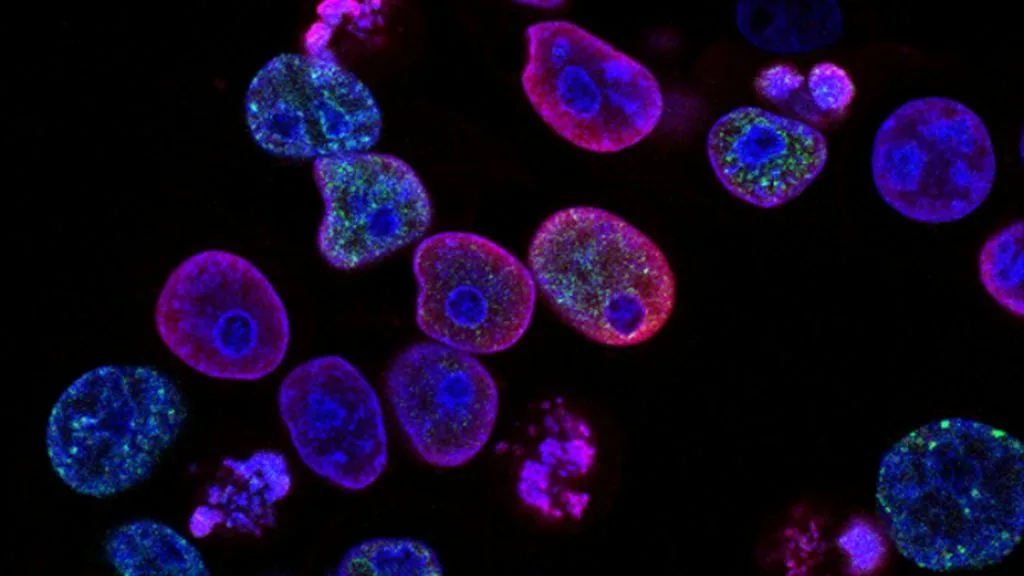
Autologous tumor cells derived directly from patient resections serve as personalized antigen sources for active specific immunotherapy and cellular vaccines. This approach exposes the host immune system to the complete patient-specific mutanome and neoantigen repertoire.
- Personalized antigen source: Autologous tumor cells capture private neoantigens unique to the individual malignancy, bypassing the limitations of shared tumor-associated antigens.
- Immune attenuation protocols: Harvested cancer cells require mechanical or enzymatic dissociation coupled with gamma-irradiation to arrest replication while preserving immunogenicity.
- Adjuvant cytokine synergy: Clinical efficacy relies on co-administration with immunostimulatory cytokines such as granulocyte-macrophage colony-stimulating factor (GM-CSF) to recruit dendritic cells.
Autologous tumor cell preparations utilize fresh surgical resection specimens or core needle biopsies harvested under sterile operating room conditions. Pathological processing requires immediate transport in cold preservation media to minimize warm ischemia time and preserve cellular viability.
The cellular immunotherapy strategy hinges on presenting the patient-specific repertoire of major histocompatibility complex (MHC) class I and class II neoepitopes to naïve T-cell repertoires. Downstream activation stimulates polyclonal cytotoxic T-lymphocyte (CTL) responses directed against private somatic mutations.
CLINICAL & LABORATORY DIAGNOSTIC PROTOCOL
- Specimen acquisition: Fresh sterile tumor tissue weighing at least 1.0 gram harvested during surgical resection, transported in cold saline with penicillin-streptomycin, and processed within 4 hours.
- Tissue dissociation: Enzymatic digestion utilizing sterile collagenase type IV and deoxyribonuclease I (DNase I) in a closed automated disaggregator system to yield a single-cell suspension.
- Viability assessment: Trypan blue dye exclusion assay confirming a minimum of 80% cellular viability prior to subsequent irradiation and cryopreservation steps.
Sterile Mechanical and Enzymatic Tissue Dissociation
Debride contaminating normal adipose and necrotic stroma from the fresh surgical specimen in a laminar flow hood. Mince tissue fragments into 1-2 millimeter cubes using sterile scalpels, transfer to digestion buffer containing collagenase (1 mg/mL) and DNase I (100 U/mL), and incubate at 37°C with continuous gentle agitation for 45 minutes.
Erythrocyte Lysis and Density Gradient Centrifugation
Filter the dissociated cell suspension through a 70-micron nylon cell strainer to remove large aggregated clumps. Centrifuge the filtrate at 400 x g for 10 minutes, resuspend the cell pellet in ACK (ammonium-chloride-potassium) lysis buffer for 3 minutes to lyse contaminating erythrocytes, and wash twice with phosphate-buffered saline.
Gamma-Irradiation Attenuation and Cryopreservation
Subject the purified single-cell autologous tumor suspension to 50 Gy to 100 Gy of cesium-137 or X-ray irradiation to induce permanent cell cycle arrest while retaining metabolic and antigenic protein synthesis. Resuspend cells in freezing medium containing 10% dimethyl sulfoxide (DMSO) and 90% autologous serum, rate-freeze in a controlled-rate freezer, and store in liquid nitrogen vapor phase.
Quality Control Microbiology and Flow Cytometry Phenotyping
Perform mandatory sterility testing for aerobic, anaerobic, and fungal pathogens using automated blood culture bottles over a 14-day incubation period. Analyze cell surface phenotype via multi-parameter flow cytometry using fluorochrome-conjugated monoclonal antibodies against CD45, EpCAM, cytokeratins, and HLA-DR to quantify tumor purity and immune cell contamination.
1. Pathophysiology, Etiology & Cellular Carcinogenesis
Autologous tumor cells harbor heterogeneous genomic aberrations driven by continuous chromosomal instability and mutational selection pressure. These malignant clones display dysregulated surface marker expression, frequent downregulation of HLA class I molecules, and evasion of immune surveillance.
Epigenetic silencing of tumor suppressor genes and aberrant DNA promoter hypermethylation further alter cellular immunogenicity and antigen processing machinery. Transcriptional repression of transporter associated with antigen processing (TAP1 and TAP2) limits endogenous peptide translocation to the endoplasmic reticulum.
The surrounding tumor microenvironment secretes immunosuppressive cytokines such as transforming growth factor-beta (TGF-beta) and interleukin-10 (IL-10). This hostile metabolic milieu inhibits dendritic cell maturation and induces functional exhaustion in tumor-infiltrating lymphocytes.
2. Oncogenic Signal Transduction Pathways & Genomic Alterations
Oncogenic activation within autologous tumor cells typically involves hyperactivation of the RAS-RAF-MEK-ERK and PI3K-AKT-mTOR intracellular signaling cascades. These dysregulated pathways promote unchecked cellular proliferation, evasion of apoptosis, and altered metabolic phenotypes characterized by aerobic glycolysis.
Downstream transcriptional alterations upregulate immune checkpoint ligands including programmed death-ligand 1 (PD-L1) on the tumor cell surface. Engagement of PD-L1 with PD-1 receptors on effector T cells triggers inhibitory intracellular signaling via SHP-1 and SHP-2 phosphatase recruitment.
Mutational burden analysis reveals diverse somatic alterations that generate private neoantigens capable of eliciting tailored cellular immune responses. High non-synonymous mutation counts correlate with increased neoepitope prediction scores and enhanced susceptibility to active specific immunotherapy.
3. Histopathological Grading, Morphological Subtypes & Differential Diagnosis
Histopathological examination of autologous tumor cell harvest tissue confirms high-grade architectural features, nuclear pleomorphism, and atypical mitotic figures. Morphological subtypes dictate processing parameters, requiring tailored enzymatic digestion protocols for dense desmoplastic stroma.
Diagnostic immunohistochemical panels utilizing cytokeratins (AE1/AE3), epithelial membrane antigen (EMA), and tissue-specific lineage markers verify tumor purity. Exclusion of non-malignant stromal elements prevents dilution of the neoplastic antigen pool during vaccine formulation.
Differential diagnosis during tissue harvesting requires careful exclusion of benign inflammatory lesions, reactive hyperplasia, and normal stromal tissue contamination. Flow cytometric sorting may be employed when primary tumor cellularity falls below acceptable diagnostic thresholds.
4. Targeted Therapeutics, Landmark Phase III Trials & Resistance Mechanics
Active specific immunotherapy utilizing autologous tumor cells is frequently combined with immune checkpoint inhibitors such as pembrolizumab or nivolumab to overcome localized immunosuppression. Monoclonal antibodies block the PD-1/PD-L1 axis, reinvigorating exhausted tumor-specific T cells primed by the cellular vaccine.
Landmark clinical investigations, including Phase III trials evaluating personalized cellular vaccines combined with granulocyte-macrophage colony-stimulating factor adjuvants, demonstrate enhanced durable progression-free survival in melanoma and renal cell carcinoma cohorts. Combination regimens consistently outperform monotherapy approaches by engaging both innate and adaptive arms.
Resistance to autologous tumor cell vaccination often arises through immunoediting, where immune selection pressures drive the outgrowth of antigen-loss variants. Upregulation of alternative checkpoint pathways, such as CTLA-3, LAG-3, and TIM-3, necessitates multi-agent blockade strategies to maintain clinical responses.
5. Molecular Staging, Minimal Residual Disease (ctDNA) & Prognostic Stratification
Integration of AJCC 8th edition TNM staging with molecular profiling refines risk stratification and identifies candidates suitable for adjuvant autologous cellular immunotherapy. High-risk resected patients with detectable minimal residual disease derive the greatest clinical benefit from active immunization protocols.
Liquid biopsy surveillance utilizing circulating tumor DNA (ctDNA) assays quantifies postoperative molecular residual disease kinetics. Clearance of ctDNA following autologous tumor cell vaccination serves as a robust surrogate marker for clinical recurrence-free survival.
Prognostic models incorporate baseline tumor mutational burden, quantitative spatial density of CD8-positive tumor-infiltrating lymphocytes, and post-vaccination delayed-type hypersensitivity skin reactions. These multidimensional biomarkers guide precision oncology clinical trial design and post-treatment surveillance intervals.