Automated Tissue Image Analysis and Deep Learning Algorithms in Cellular Pathology: Diagnostic Algorithmic Validation, Morphometric Feature Extraction & Quality Control Standards
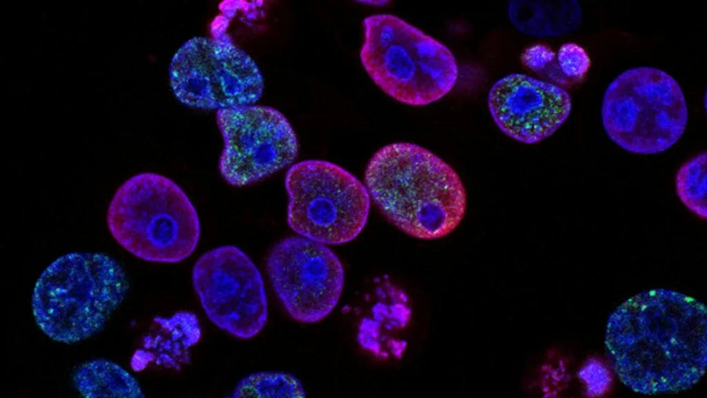
Automated tissue image analysis applies convolutional neural networks and advanced computer vision to digital whole slide images, enabling quantitative morphometric profiling and biomarker quantification in oncologic pathology. Standardization of digital workflows requires rigorous calibration of slide scanners, color normalization protocols, and validation against established human observer benchmarks.
- Digital whole slide imaging requires high-resolution scanning at 40x magnification with strict focus plane validation.
- Convolutional neural networks achieve high concordance with expert pathologists in quantifying nuclear atypia, mitotic figures, and immunohistochemical staining intensity.
- Algorithmic scoring minimizes inter-observer variability in predictive biomarker assays such as HER2 dual-probe ISH and PD-L1 immunohistochemistry.
Digital pathology implementation demands rigorous end-to-end scanner validation and standardized color constancy across heterogeneous tissue staining batches. High-resolution whole slide imaging scanners capture gigapixel histological files that serve as the primary input matrix for computational vision algorithms.
Deep learning architectures, specifically U-Net segmentation models and ResNet classification backbones, process digitized tissue sections to segment epithelial compartments, stromal elements, and infiltrating immune populations. These computational pipelines execute automated morphometric feature extraction to derive prognostic risk scores from routine hematoxylin and eosin preparations.
CLINICAL & LABORATORY DIAGNOSTIC PROTOCOL
- Scanner calibration: Daily scanner calibration using NIST-traceable micrometers and color calibration targets to ensure consistent RGB spectral acquisition.
- Image compression: Lossless or high-quality wavelet compression (e.g., JPEG 2000) configured to preserve sub-micron nuclear membrane details without compression artifact interference.
- Validation cohort: Minimum 60 clinical glass slides spanning the full spectrum of diagnostic difficulty evaluated independently by three board-certified pathologists to establish ground truth.
Whole Slide Image Acquisition & Quality Control
Scan stained glass slides at 40x optical magnification (0.25 microns per pixel resolution) utilizing automated focus tracking across multiple focal planes. Inspect acquired whole slide images for out-of-focus blurring, tiling artifacts, dust particles, and glass coverslip air bubbles.
Color Normalization & Tissue Background Segmentation
Apply Macenko or Reinhard color normalization algorithms to standardize hematoxylin and DAB stain density across different staining lots and laboratory sites. Execute background thresholding via Otsu's method to separate diagnostic tissue pixels from glass slide white space.
Deep Learning Object Detection & Nuclei Segmentation
Deploy validated convolutional neural networks trained on multi-institutional datasets to segment individual cell nuclei and classify cellular phenotypes (tumor cells, lymphocytes, stromal fibroblasts, endothelial cells). Calculate spatial distribution maps and nuclear-to-cytoplasmic ratios.
Biomarker Quantification & Automated Scoring Reporting
Execute targeted scoring algorithms to compute immunohistochemical positivity metrics, including Ki-67 proliferation index percentages and HER2 amplification ratios. Generate structured digital pathology reports incorporating heatmaps of marker expression heterogeneity.
1. Computational Architecture of Digital Pathology Systems
Digital whole slide imaging systems utilize high-aperture optics and automated stage encoders to capture microscopic fields and stitch them into gigapixel raster files. These files exceed standard image viewing capacities, necessitating pyramidal multi-resolution file formats such as DICOM, SVS, or NDPI for efficient computational streaming.
Convolutional neural networks process these high-resolution tiles through hierarchical feature extraction layers, identifying low-level edges in initial convolutional filters and assembling high-level semantic tissue architectures in deeper layers. Spatial attention mechanisms allow algorithms to focus computational power on diagnostically dense areas while ignoring uninformative stromal background.
Hardware acceleration via graphics processing units is essential for executing real-time inference on large cohorts of oncologic tissue sections. Cloud-based distributed computing frameworks manage data transfer, load balancing, and secure storage compliance in accordance with institutional data protection regulations.
2. Morphometric Feature Extraction and Nuclear Phenotyping
Automated nuclear segmentation isolates individual cancer cell nuclei to quantify morphometric features including nuclear area, perimeter, eccentricity, major-to-minor axis ratio, and chromatin texture coarseness. These quantitative descriptors correlate strongly with nuclear grade and biological aggressiveness across diverse solid malignancies.
Graph neural networks model the spatial relationships between segmented tumor cells and tumor-infiltrating lymphocytes by constructing cellular adjacency graphs. Topological data analysis of these graphs extracts quantitative metrics of immune infiltration patterns, distinguishing between 'inflamed', 'excluded', and 'desert' tumor microenvironments.
Feature selection algorithms eliminate redundant radiomic and morphometric variables to prevent overfitting, retaining a robust panel of phenotypic signatures predictive of recurrence and metastatic potential. These computational phenotypes augment traditional subjective grading systems with reproducible continuous numerical scores.
3. Algorithmic Standardization of Predictive Biomarkers
Immunohistochemical assays such as programmed death-ligand 1 (PD-L1) evaluation in non-small cell lung carcinoma suffer from inter-observer variability driven by subjective scoring of tumor proportion scores. Automated tissue image analysis applies standardized pixel intensity thresholds to distinguish true membranous DAB chromogen deposition from cytoplasmic background and edge artifacts.
Human epidermal growth factor receptor 2 (HER2) quantification via brightfield in situ hybridization or immunohistochemistry benefits from automated signal counting algorithms that eliminate counting bias in heterogeneous breast carcinoma specimens. Algorithms accurately differentiate clustered gene amplification signals from single copynumber variants within individual nuclei.
Quality assurance protocols continuously monitor algorithm performance by tracking algorithmic drift, batch staining variations, and scanner optical degradation. Regular validation against reference standard cohorts ensures clinical safety and regulatory compliance under College of American Pathologists standards.
4. Validation Methodologies and Clinical Workflow Integration
Clinical validation of computational pathology tools requires comprehensive concordance studies measuring overall agreement, positive percent agreement, and negative percent agreement against consensus human reference standards. Studies must encompass challenging edge-case specimens including necrotic cores, extensive cautery artifact, and low-cellularity core needle biopsies.
Integration of automated tissue image analysis into laboratory information systems requires HL7 and FHIR interoperability standards to transmit structured algorithmic scores directly into electronic health records. Pathologist workstations display diagnostic heatmaps that overlay algorithm outputs onto whole slide images for rapid visual verification.
Regulatory clearance pathways, including FDA 510(k) clearance and CE-IVDR certification, mandate rigorous post-market surveillance to verify algorithm generalizability across different tissue fixatives, tissue microarrays, and staining platforms used in global clinical trials.
5. Future Directions: Multimodal AI and Spatial Transcriptomics
Emerging computational frameworks integrate digital histology whole slide images directly with genomic sequencing data, transcriptomic profiles, and multiplex immunofluorescence to construct comprehensive spatial atlases of tumors. Multimodal deep learning models leverage these combined inputs to predict specific driver mutations directly from hematoxylin and eosin morphology.
Spatial transcriptomics technologies generate high-plex gene expression matrices mapped to exact microscopic tissue coordinates, requiring advanced computational algorithms to align molecular data with digital histology images. These joint analytical pipelines uncover novel cellular neighborhoods and intercellular signaling networks driving therapeutic resistance.
Continuous active learning paradigms allow deployed algorithms to query human pathologists on ambiguous edge cases, updating neural network weights securely to improve diagnostic accuracy over time without catastrophic forgetting of established pathological patterns.