BRAF V600E and V600K Oncogenic Mutations in Melanoma and Colorectal Tumors: Molecular Pathology, Inhibitor Resistance, and Dual-Targeted Therapeutics
Activating **BRAF V600E** and **V600K** mutations drive constitutional kinase activation within the **RAS-RAF-MEK-ERK** signaling cascade across malignant melanoma and colorectal carcinomas. Precision combinatorial therapies targeting mutant kinase complexes have fundamentally reshaped clinical survival paradigms.
- Primary molecular driver: Substitution of valine at codon 600 with glutamic acid (V600E) or lysine (V600K) within the kinase domain of **BRAF**.
- First-line standard of care in melanoma: Combination **dabrafenib** plus **trametinib** or **encorafenib** plus **binimetinib**.
- Primary mechanism of resistance: Reactivation of the **MAPK** pathway via upstream **RTK** upregulation or downstream **MEK1/2** secondary mutations.
Cutaneous melanomas harbor **BRAF** mutations in approximately 40 to 50 percent of cases, with **V600E** accounting for approximately 80 percent of these alterations. Conversely, metastatic colorectal carcinomas exhibit **BRAF V600E** mutations in roughly 8 to 12 percent of patients, frequently correlating with microsatellite instability and advanced age.
The replacement of valine at codon 600 locks the **BRAF** kinase domain into an active conformation independent of upstream **RAS** GTPase activation. This constitutional enzymatic state hyperactivates downstream **MEK1/2** and **ERK1/2** phosphorylation cascades, driving uncontrolled cellular proliferation and survival.
CLINICAL & LABORATORY DIAGNOSTIC PROTOCOL
- Specimen acquisition: Formalin-fixed, paraffin-embedded (FFPE) tissue blocks containing a minimum of 20 percent neoplastic cellularity derived from core biopsies or surgical resections.
- Tissue processing: Fixation in 10 percent neutral-buffered formalin for 6 to 72 hours, processed via automated dehydrators, and embedded in paraffin wax blocks.
- Control standards: Wild-type and known **BRAF V600E** mutant cell line controls processed in parallel with diagnostic analytical runs to verify assay sensitivity.
Genomic DNA Extraction & Quantification
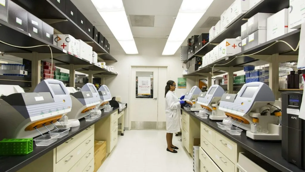
Microdissect tumor-enriched regions from 5-micron unstained FFPE sections using H and E-guided manual scraping. Extract genomic DNA utilizing silica-column magnetic bead chemistry and quantify double-stranded DNA using fluorometric assays.
Multiplex PCR Amplification & NGS Library Construction
Amplify target exons harboring codon 600 of **BRAF** using target-specific multiplex primer pools. Prepare sequencing libraries by ligating Illumina-compatible adapters with unique dual indexes during enzymatic end-repair.
Massively Parallel Sequencing & Variant Calling
Load denatured libraries onto a sequencing-by-synthesis platform (e.g., Illumina MiSeq or NextSeq 550 Dx) to achieve a minimum targeted sequencing depth of 500x coverage. Execute bioinformatics alignment against the hg19/GRCh37 reference genome.
Orthogonal Validation via Immunohistochemistry (IHC)
Perform automated immunohistochemical staining using the anti-**BRAF V600E** mouse monoclonal primary antibody clone VE1. Visualize immune complexes with horseradish peroxidase polymer detection and DAB chromogen substrate.
1. Pathophysiology, Etiology & Cellular Carcinogenesis
Oncogenic activation of **BRAF** triggers sustained transcriptional upregulation of Cyclin D1, facilitating unregulated G1-to-S phase cell cycle progression. Chronic pathway engagement evades cellular senescence checkpoints and impairs apoptotic signaling via transcriptional repression of pro-apoptotic **BCL-2** family proteins.
Epigenetic dysregulation in **BRAF** mutant colorectal tumors frequently manifests as the CpG island methylator phenotype (CIMP-high). This hypermethylated state silences the mismatch repair gene **MLH1**, resulting in microsatellite instability and distinct clinical behaviors.
The tumor microenvironment in **BRAF**-mutated malignancies exhibits variable immune infiltration characterized by dense immunosuppressive myeloid cells and regulatory T-cell networks. Upregulation of **PD-L1** expression driven by constitutive **MAPK** signaling provides an immune evasion mechanism.
2. Oncogenic Signal Transduction Pathways & Genomic Alterations
Wild-type **BRAF** functions as a cytoplasmic serine/threonine kinase regulated by transient **RAS**-GTP binding at the inner plasma membrane. Mutational substitution at codon 600 mimics electrostatic phosphorylation, stabilizing the active kinase conformation without upstream signaling.
Constitutively active **BRAF** phosphorylates downstream **MEK1** and **MEK2**, which subsequently activate **ERK1** and **ERK2** kinases via dual phosphorylation of threonine and tyrosine residues. Translocated nuclear **ERK** phosphorylates multiple transcription factors including **c-Fos**, **c-Jun**, and **MYC**.
Resistance mechanisms frequently bypass mutant **BRAF** inhibition through parallel activation of the **PI3K-AKT-mTOR** pathway or receptor tyrosine kinase amplification. Feedback loop reactivation via **EGFR** upregulation is particularly prominent in colorectal cancers treated with selective inhibitors.
3. Histopathological Grading, Morphological Subtypes & Differential Diagnosis
Cutaneous melanomas harboring **BRAF V600** mutations frequently present with nodular or superficial spreading architectures characterized by nested melanocytic proliferation and cytologic atypia. Colorectal carcinomas with this driver often display poorly differentiated, mucinous, or serrated glandular morphology with abundant inflammatory infiltrates.
Diagnostic immunohistochemistry panels differentiate primary sites and mutation subtypes utilizing markers such as **SOX10**, **S100**, **CK20**, and **CDX2**. The **VE1** mutation-specific monoclonal antibody serves as a reliable surrogate for molecular testing in specialized pathology laboratories.
Differential diagnosis requires exclusion of wild-type melanomas, non-V600 **BRAF** non-canonical alterations, and metastatic adenocarcinomas of alternative primary origins. Definitive confirmation mandates multiplex molecular assays to guide therapeutic selection.
4. Targeted Therapeutics, Landmark Phase III Trials & Resistance Mechanics
First-generation mutant-specific kinase inhibitors including **dabrafenib** and **vemurafenib** revolutionized systemic therapy for advanced cutaneous melanoma. The addition of **MEK** inhibitors such as **trametinib** or **cobimetinib** mitigated paradoxical **MAPK** activation and delayed acquired resistance.
The landmark Phase III **COMBI-d** and **COMBI-v** clinical trials established **dabrafenib** plus **trametinib** combination therapy as a standard of care, demonstrating superior progression-free survival and overall survival compared to monotherapy. In colorectal cancer, the Phase III **BEACON CRC** trial proved the superiority of **encorafenib** combined with **cetuximab** over standard chemotherapy.
Acquired resistance mechanisms encompass secondary **NRAS** or **MEK1** mutations, **BRAF** amplification, and activation of alternative receptor tyrosine kinases. Novel investigative regimens evaluate fourth-generation pan-RAF inhibitors and multikinase inhibitors to overcome these tertiary escape routes.
5. Molecular Staging, Minimal Residual Disease (ctDNA) & Prognostic Stratification
Integration of **BRAF** mutation status with AJCC 8th edition TNM staging refines prognostic stratification for cutaneous melanoma and colorectal carcinoma patients. Colorectal tumors harboring **BRAF V600E** mutations consistently demonstrate poorer overall survival compared to wild-type counterparts within equivalent TNM stages.
Circulating tumor DNA (ctDNA) liquid biopsy assays enable non-invasive surveillance of minimal residual disease and early detection of treatment resistance mutations. Serial plasma monitoring permits quantitative tracking of **BRAF** variant allele frequencies during targeted therapy administration.
Prognostic models increasingly incorporate genomic signatures, tumor mutation burden, and microenvironment markers to optimize therapeutic sequencing. Prospective clinical trials continue to evaluate ctDNA-guided treatment escalations in resected stages of disease.