Cancer Pain: Neuro-Oncological Mechanisms, Somatosensory Pathway Transduction, and Multimodal Pharmacological Management
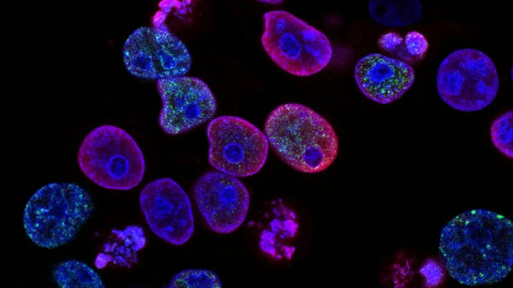
Cancer pain is a complex multifactorial symptom affecting over 70% of patients with advanced malignancies, driven by direct tumor infiltration, nerve compression, and treatment-related toxicities. Its pathophysiology requires precise neurobiological evaluation to target peripheral nociceptors and central sensitization pathways.
- Primary mechanistic driver: Ectopic firing of damaged primary afferent C-fibres and A-delta fibres via upregulation of voltage-gated sodium channels.
- First-line pharmacological standard of care: Step 2 and Step 3 opioids on the WHO analgesic ladder combined with structured adjuvant co-analgesics.
- Primary therapeutic challenge: Management of neuropathic cancer pain resistant to standard opioid titration, necessitating neuromodulatory and interventional modalities.
Cancer pain manifests across varying clinical stages, demonstrating a high prevalence in patients with metastatic bone lesions, perineural invasion, and hollow viscus obstruction. Epidemiological investigations confirm that somatic, visceral, and neuropathic etiologies frequently coexist, requiring structured diagnostic stratification to optimize clinical outcomes.
The cellular pathogenesis involves continuous release of inflammatory cytokines, chemokines, and growth factors within the tumor microenvironment that sensitize peripheral nociceptors. This persistent local stimulation drives sustained depolarization of dorsal root ganglion neurons and promotes spinal cord dorsal horn central sensitization.
CLINICAL & LABORATORY DIAGNOSTIC PROTOCOL
- Clinical pain assessment: Comprehensive multidomain evaluation utilizing the Brief Pain Inventory (BPI) and Edmonton Symptom Assessment System (ESAS) to quantify baseline intensity.
- Diagnostic imaging: High-resolution contrast-enhanced magnetic resonance imaging (MRI) of the neuraxis or computed tomography (CT) to delineate structural nerve root compression or osseous lysis.
- Laboratory validation: Serum complete blood count, comprehensive metabolic panel, and inflammatory markers (C-reactive protein) to evaluate systemic contributory factors.
Comprehensive Pain Characterization and Somatosensory Mapping
Administer standardized validated questionnaires including the Douleur Neuropathique 4 (DN4) questionnaire to differentiate somatic, visceral, and neuropathic pain components. Map dermatomal boundaries, sensory deficits, and mechanical allodynia using standardized Semmes-Weinstein monofilaments and thermal probes.
Radiological Localization of Oncologic Structural Etiology
Perform targeted multi-planar neuroimaging (MRI spine or brain with gadolinium contrast) to evaluate leptomeningeal disease, epidural spinal cord compression, or perineural tumor spread. Identify exact vertebral body involvement and structural compromise utilizing established Tomita or Tokuhashi scoring systems.
Multimodal Pharmacological Titration and WHO Ladder Integration
Initiate non-opioid analgesics (acetaminophen, NSAIDs if contraindicated) for mild somatic pain, escalating immediately to WHO Step 3 opioid agonists (morphine, oxycodone, hydromorphone) for moderate-to-severe nociceptive pain. Administer adjuvant neuropathic agents (gabapentinoids, serotonin-norepinephrine reuptake inhibitors) for confirmed neuropathic pain syndromes.
Interventional Procedural Evaluation and Multidisciplinary Review
Assess patient eligibility for image-guided interventional procedures, including fluoroscopy-guided celiac plexus neurolysis for upper abdominal visceral pain or targeted epidural steroid injections. Convene multidisciplinary tumor board review to evaluate palliative radiotherapy, surgical decompression, or intrathecal drug delivery system implantation.
1. Pathophysiology, Etiology & Cellular Carcinogenesis
Malignant neoplasms induce tissue damage through direct mechanical compression, infiltration of bone periosteum, and release of osteolytic factors such as **parathyroid hormone-related protein (PTHrP)**. This osteolytic cascade activates osteoclasts, leading to structural microfractures and localized acidosis that stimulates acid-sensing ion channels on sensory nerve terminals.
Perineural invasion represents a distinct pathological mechanism wherein carcinoma tracking along nerve sheaths secretes neurotrophic factors including **nerve growth factor (NGF)** and **brain-derived neurotrophic factor (BDNF)**. These ligands bind to high-affinity tyrosine kinase receptors on primary afferent neurons, driving axonal remodeling and spontaneous ectopic discharges.
The tumor microenvironment functions as an active neuroimmune interface containing infiltrating macrophages, T-lymphocytes, and mast cells that secrete **interleukin-6 (IL-6)** and **tumor necrosis factor-alpha (TNF-alpha)**. These pro-inflammatory cytokines lower the activation threshold of peripheral nociceptors and recruit additional inflammatory cells to perpetuate chronic hyperexcitability.
2. Oncogenic Signal Transduction Pathways & Genomic Alterations
At the molecular level, peripheral nerve injury and tumor-derived mediators trigger the upregulation of voltage-gated sodium channels **Nav1.7**, **Nav1.8**, and **Nav1.9** along dorsal root ganglion neurons. This channelopathy lowers the resting membrane potential, facilitating spontaneous generation of action potentials and hyperalgesia.
Intracellular nociceptive signaling relies heavily on the **protein kinase A (PKA)** and **protein kinase C (PKC)** intracellular cascades, which phosphorylate peripheral receptor proteins like **TRPV1**. Persistent pathway activation results in receptor sensitization and enhanced calcium influx into the primary sensory neuron terminal.
Central sensitization within the spinal cord dorsal horn is mediated by sustained release of **glutamate** binding to **N-methyl-D-aspartate (NMDA)** receptors. This persistent activation removes magnesium blockade, promoting intracellular calcium accumulation and long-term synaptic potentiation in second-order projection neurons.
3. Histopathological Grading, Morphological Subtypes & Differential Diagnosis
Histopathological examination of biopsied tumor tissue reveals distinct architectural growth patterns, such as perineural infiltration characterized by malignant cell clusters surrounding neural epineurium. Special stains and immunohistochemical panels including **S100 protein** and **glial fibrillary acidic protein (GFAP)** confirm the extent of nerve bundle destruction.
Differential diagnosis of cancer-induced pain requires distinguishing malignant neuropathy from treatment-induced toxicities such as chemotherapy-induced peripheral neuropathy (**CIPN**). Pathological evaluation of nerve biopsies in CIPN demonstrates axonal degeneration, mitochondrial swelling, and localized microtubule disruption.
Accurate pathological classification also differentiates tumor-induced bone destruction from metabolic bone diseases, utilizing immunohistochemical markers such as **RUNX2** and **RANKL** expression profiles. This histological precision ensures clinicians distinguish direct metastatic invasion from osteoporotic or radiation-induced insufficiency fractures.
4. Targeted Therapeutics, Landmark Phase III Trials & Resistance Mechanics
Pharmacological management utilizes strong opioid agonists including **morphine sulfate**, **oxycodone hydrochloride**, and **hydromorphone**, acting on central and peripheral **mu-opioid receptors (OPRM1)**. Landmark clinical investigations, including comparative trials evaluated by the World Health Organization, validate systematic titration protocols for achieving rapid analgesia.
For neuropathic pain components, phase-proven adjuvant medications such as **gabapentin** and **pregabalin** bind to the alpha-2-delta subunit of voltage-gated calcium channels, reducing presynaptic neurotransmitter release in the dorsal horn. Randomized controlled trials demonstrate significant reductions in daily pain scores and improved quality of life metrics compared to placebo.
Refractory cancer pain frequently develops tolerance or opioid-induced hyperalgesia driven by **N-methyl-D-aspartate (NMDA)** receptor upregulation and descending facilitation. Salvage strategies incorporate multimodal adjuvant administration, **ketamine** infusions for NMDA antagonism, and surgical neuromodulation via intrathecal drug delivery systems.
5. Molecular Staging, Minimal Residual Disease (ctDNA) & Prognostic Stratification
Prognostic stratification of patients with severe cancer pain integrates the **Edmonton Classification of Cancer Pain (ECCP)** with traditional AJCC TNM staging parameters. Patients exhibiting neuropathic or mixed pain mechanisms accompanied by high psychological distress demonstrate significantly reduced overall survival and treatment tolerance.
Advanced liquid biopsy surveillance utilizing **circulating tumor DNA (ctDNA)** quantifies minimal residual disease and tracks molecular recurrence prior to overt radiological progression. Monitoring somatic mutation allele frequencies enables early detection of treatment failure and adjustment of palliative strategies.
Integrating molecular staging biomarkers with patient-reported pain outcomes establishes a robust predictive framework for oncologic care. Prospective clinical paradigms utilize these comprehensive datasets to personalize interventional pain interventions and improve end-of-life quality metrics.