Cellular Friability in Malignant Neoplasia: Histological Artifacts, Diagnostic Pitfalls, and Matrix Metalloproteinase Dysregulation
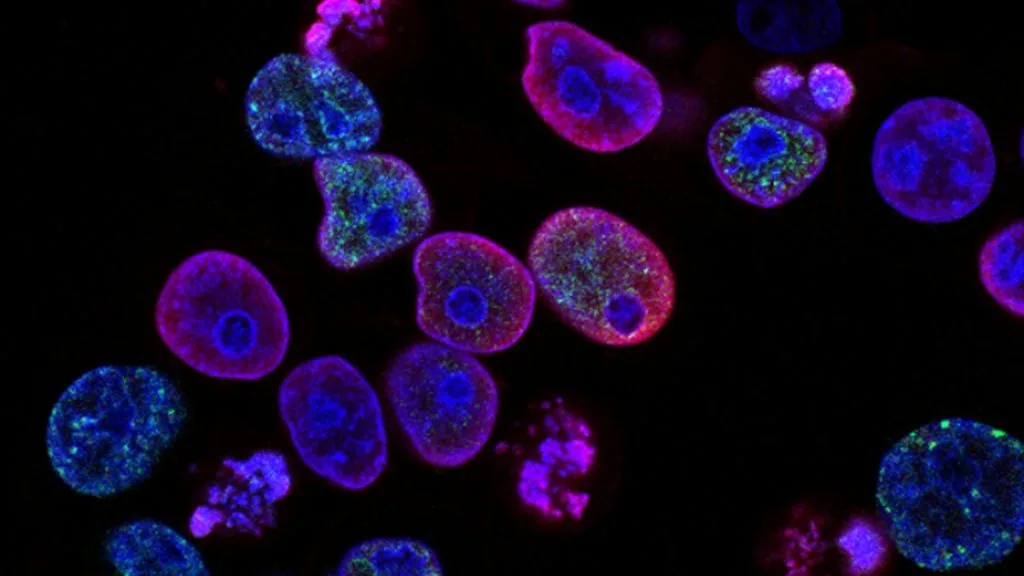
Cellular friability in clinical histopathology denotes an inherent tissue fragility characterized by extreme susceptibility to mechanical fragmentation during biopsy acquisition and slide processing. This physical manifestation correlates directly with extracellular matrix degradation and proteolytic enzyme overexpression in invasive neoplasms.
- Mechanical definition: Tissue friability creates prominent crush artifacts, nuclear streaming, and tissue disintegration during microtome sectioning.
- Biochemical driver: Upregulation of matrix metalloproteinases (MMP-1, MMP-9) degrades collagenous stromal scaffolds.
- Diagnostic consequence: Excessive friability frequently masquerades as fixation artifact, obscuring true architectural atypia.
Cellular friability presents diagnostic challenges across diverse oncologic subspecialties, particularly when procuring core needle biopsies from hypervascular, poorly cohesive carcinomas. Pathologists frequently observe disrupted cellular cohesion in high-grade urothelial carcinomas, neuroendocrine neoplasms, and poorly differentiated adenocarcinomas.
The cellular mechanism underlying friability involves the proteolytic cleavage of cell-adhesion molecules and structural stromal proteins. Disruption of the E-cadherin-catenin complex at adherens junctions leads to diminished intercellular adhesion and increased mechanical fragility.
CLINICAL & LABORATORY DIAGNOSTIC PROTOCOL
- Specimen acquisition: Biopsy specimens handled with atraumatic forceps to minimize mechanical shearing of friable tumor nests.
- Tissue fixation: Immediate immersion in 10% neutral-buffered formalin for exactly 24 hours to stabilize fragile nuclear membranes.
- Control standards: Internal normal stromal elements utilized as baseline references for evaluating fixation artifact versus true mechanical fragility.
Gentle Biopsy Processing & Embedding
Transfer friable core biopsies into biopsy mesh cassettes immediately. Avoid excessive mechanical manipulation during paraffin embedding to prevent fragmentation.
Cryo-Sectioning & Microtomy Calibration
Chill paraffin blocks on ice plates for 15 minutes prior to sectioning. Set microtome clearance angle to 5 to 7 degrees and section at 3-micron thickness.
Adhesion Slide Mounting & HIER
Mount sections on positively charged silanized glass slides. Perform heat-induced epitope retrieval in pH 9.0 Tris-EDTA buffer at 98 degrees Celsius for 20 minutes.
Immunohistochemical Staining & Evaluation
Apply primary monoclonal antibodies against E-cadherin and MMP-9 using an automated stainer. Counterstain with modified Harris hematoxylin.
1. Pathophysiology, Etiology & Extracellular Matrix Degradation
Tissue friability arises from the structural collapse of the tumor microenvironment due to the enzymatic destruction of interstitial collagen. Tumor-associated macrophages and carcinoma cells secrete high levels of matrix metalloproteinases that cleave type I and type III collagens.
Epigenetic silencing of cell adhesion genes compounds this structural vulnerability by abolishing desmosomal and adherens junction formation. Consequently, neoplastic cells lose mechanical integrity and shear apart under standard diagnostic processing forces.
The surrounding desmoplastic stroma often exhibits hyalinization and myxoid degeneration, creating a fragile matrix that fails to anchor epithelial cell islands. This breakdown produces the classic friable architecture observed in endoscopic and surgical pathology.
2. Proteolytic Cascades and Cell-Adhesion Disruption
The molecular basis of friability centers on the aberrant activation of the plasminogen activator system and the MMP cascade. Activation of MMP-2 and MMP-9 degrades basement membrane laminin and type IV collagen, stripping epithelial nests of structural support.
Concurrently, loss of membranous **CDH1** expression eliminates homotypic cell-cell binding mediated by E-cadherin. Intracellular signaling shifts toward pathways that promote cellular dissociation, such as the Rho-associated protein kinase pathway.
These biochemical alterations transform cohesive epithelial sheets into fragile, dyshesive cell populations. The resulting tissue behaves as a semi-fluid mass that readily fragments during surgical resection.
3. Histopathological Artifacts and Diagnostic Differentiation
Pathologists must differentiate between true biological friability and procedural crush artifact induced by dull microtome blades or rough forceps handling. True friability typically features dyscohesive single cells embedded within a markedly degraded, myxoid stroma.
Crush artifact presents with nuclear streaming, hyperchromatic smudging, and loss of cytoplasmic detail without underlying enzymatic matrix destruction. Diagnostic distinction is critical to avoid misinterpreting fragile artifactual debris as invasive micropapillary carcinoma.
Immunohistochemical panels utilizing **CKAE1/AE3** and **Vimentin** assist in confirming epithelial differentiation within fragmented, friable biopsy specimens. Internal controls help verify that architectural disruption is intrinsic to the neoplasm rather than secondary to handling.
4. Clinical Implications, Surgical Challenges and Therapeutics
Surgical management of friable neoplasms requires meticulous technique, as fragile tumors rupture easily during oncologic resection, spilling malignant cells. Intraoperative fragmentation increases the risk of positive surgical margins and local peritoneal or pleural dissemination.
Landmark surgical trials emphasize the necessity of en-bloc resection without capsule violation, even when dealing with friable tumor consistency. Pharmacological inhibition of extracellular matrix remodeling enzymes remains an area of active clinical investigation.
Combined systemic regimens utilizing platinum agents combined with anti-angiogenic therapies aim to stabilize the tumor vasculature and reduce matrix turnover. Clinical monitoring via circulating tumor DNA assays provides complementary prognostic data when tissue biopsies are severely fragmented.
5. Molecular Staging, Prognostic Stratification and Biomarkers
The presence of pronounced cellular friability correlates with aggressive tumor behavior, advanced **AJCC 8th Edition** staging, and high metastatic potential. High expression levels of **MMP9** and **COL1A1** serve as molecular indicators of aggressive extracellular breakdown.
Pathologists incorporate tissue architecture and friability assessments into synoptic reporting to guide oncology teams regarding local recurrence risks. Standardized grading ensures reproducibility across academic medical centers and clinical trials.
Future diagnostic paradigms will integrate artificial intelligence algorithms trained on digital whole-slide images to quantify tissue fragmentation indices automatically. This computational approach aims to standardize the assessment of friability in routine oncologic pathology.