Circulating Tumor DNA and Plasma Cancer Likelihood: Liquid Biopsy Kinetics, Analytical Validity & Oncologic Diagnostics
Cancer likelihood in plasma evaluates the quantitative and qualitative presence of circulating cell-free tumor DNA shed from primary and metastatic neoplasms into the vascular compartment. This diagnostic paradigm provides high sensitivity for somatic mutation profiling, minimal residual disease tracking, and therapeutic resistance monitoring.
- Circulating tumor DNA fraction correlates directly with tumor volume, vascularity, cellular turnover, and metastatic burden.
- Next-generation sequencing (NGS) panels detect actionable somatic mutations in plasma with high concordance to tissue biopsies.
- Serial plasma sampling identifies secondary resistance mutations months before radiographic disease progression occurs.
Cell-free DNA circulates in healthy individuals primarily as a consequence of physiological apoptosis and hematopoietic cell turnover, yielding low baseline plasma concentrations. In contrast, patients with malignant neoplasms exhibit elevated plasma cfDNA concentrations driven by necrotic cell lysis, active cellular secretion, and macrophage phagocytosis of tumor tissue.
The fraction of cfDNA derived from neoplastic clones, designated as circulating tumor DNA, harbors identical somatic alterations, chromosomal aberrations, and epigenetic signatures found in the corresponding solid tumor. Analytical quantification of this tumor-derived fraction enables non-invasive detection of cancer likelihood and molecular profiling when tissue acquisition is clinically unfeasible.
CLINICAL & LABORATORY DIAGNOSTIC PROTOCOL
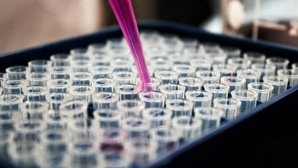
- Specimen acquisition: Collect 10 mL of whole blood into specialized cell-free DNA collection tubes (Streck Cell-Free DNA BCT) containing a formaldehyde-free preservative to prevent leukocyte lysis and genomic DNA contamination.
- Sample processing: Centrifuge whole blood at 1,600 x g for 10 minutes at room temperature within 2 hours of phlebotomy to isolate plasma, followed by a high-speed secondary spin (16,000 x g for 10 minutes) to eliminate cellular debris.
- Control standards: Extract total circulating nucleic acids using automated magnetic bead-based assays with internal synthetic spike-in controls to monitor extraction efficiency and library preparation recovery.
Cell-Free DNA Extraction & Quantification
Isolate cfDNA from 2 to 4 mL of clarified plasma using silica-membrane or magnetic bead columns optimized for low-molecular-weight fragments (100–200 base pairs). Quantify extracted nucleic acids using high-sensitivity fluorescent dyes (Qubit dsDNA HS Assay) and assess fragment size distribution via automated capillary electrophoresis (Agilent TapeStation 4200).
Targeted Library Preparation & Molecular Barcoding
Construct sequencing libraries using 10 to 50 nanograms of extracted cfDNA utilizing unique molecular identifiers (UMIs) ligated to DNA fragments prior to amplification. Apply targeted hybrid-capture panels directed against canonical cancer driver genes (**TP53**, **KRAS**, **EGFR**, **PIK3CA**) to enrich tumor-specific sequences.
Massively Parallel Sequencing (NGS)
Perform ultra-deep sequencing on a designated high-throughput platform (Illumina NovaSeq 6000 or equivalent) achieving a mean target sequencing depth exceeding 10,000x unique coverage. Sequence generated clusters using paired-end chemistry with 2 x 150 bp read lengths to capture complete cfDNA insert sizes.
Bioinformatic Pipeline Analysis & Variant Calling
Execute secondary and tertiary bioinformatic pipelines incorporating UMI consensus error suppression, alignment to the GRCh38 human reference genome, and somatic variant calling via specialized algorithms. Filter variants against population databases (gnomAD) and clonal hematopoiesis of indeterminate potential (CHIP) filter lists to report true tumor variants.
1. Pathophysiology, Etiology & Cellular Carcinogenesis
Malignant transformation initiates through stochastic genetic mutations and chromosomal instability within epithelial, mesenchymal, or hematopoietic stem cell compartments. As nascent tumors expand beyond a critical diffusion limit of 1 to 2 millimeters, regions of cellular hypoxia develop, driving upregulation of hypoxia-inducible factor 1-alpha and subsequent neoangiogenesis.
The resulting tumor vasculature is structurally aberrant, characterized by disorganized endothelial lining, excessive pericyte detachment, and elevated interstitial fluid pressure. These vascular abnormalities promote cellular shedding, necrosis, and active release of nucleic acid fragments into the systemic circulation, establishing the biological basis for plasma cancer detection.
Epigenetic dysregulation, encompassing global DNA hypomethylation and site-specific promoter hypermethylation of tumor suppressor genes, further contributes to aberrant transcriptional profiles and distinctive cfDNA methylation patterns. The tumor microenvironment actively secretes exosomes containing double-stranded DNA, RNA, and protein cargo that circulate alongside naked cell-free DNA fragments.
2. Oncogenic Signal Transduction Pathways & Genomic Alterations
Oncogenic activation frequently converges upon key regulatory networks, including the **RAS-RAF-MEK-ERK** and **PI3K-AKT-mTOR** intracellular signaling cascades. Activating mutations within **KRAS** codons 12 and 13 or **BRAF** V600E drive constitutive kinase activity, promoting unchecked cellular proliferation, evasion of apoptosis, and metabolic reprogramming.
Receptor tyrosine kinases such as **EGFR**, **ERBB2**, and **MET** undergo ligand-independent dimerization, autophosphorylation, and conformational shifts that hyperactivate downstream transcriptional programs. In parallel, inactivation of gatekeeper tumor suppressors like **TP53** abrogates the G1/S cell cycle checkpoint, permitting genomic instability and survival under genotoxic stress.
Targeted therapeutic blockade exerts selective pressure on these signaling pathways, frequently engendering secondary acquired resistance mutations. Detection of emerging genomic alterations in plasma provides direct insight into evolving tumor subclones without necessitating repeat surgical biopsies.
3. Histopathological Grading, Morphological Subtypes & Differential Diagnosis
While plasma assays evaluate molecular profiles rather than architectural histology, circulating tumor DNA concentrations correlate strongly with histological grade, tumor cellularity, and mitotic activity. High-grade carcinomas with solid growth patterns and extensive geographic necrosis consistently shed higher quantities of cfDNA than low-grade, well-differentiated neoplasms.
Diagnostic validation requires correlating plasma somatic findings with tissue histopathology and immunohistochemical staining panels. For pulmonary neoplasms, confirmation of primary tumor origin relies on positive expression of **TTF-1** and **Napsin A** for adenocarcinomas, or **p40** and **CK5/6** for squamous cell carcinomas.
Differential diagnosis of elevated plasma mutant allele frequency must exclude benign hematological conditions, inflammatory states, and clonal hematopoiesis of indeterminate potential. Somatic mutations identified in **DNMT3A**, **TET2**, and **ASXL1** typically originate from aging hematopoietic stem cells rather than solid tumors and require careful bioinformatic filtration.
4. Targeted Therapeutics, Landmark Phase III Trials & Resistance Mechanics
Precision oncology relies on matching specific genomic alterations with targeted pharmacological inhibitors designed to disrupt oncogenic signaling pathways. First- and second-generation tyrosine kinase inhibitors have been largely superseded by third-generation agents like **osimertinib**, which irreversibly binds mutant **EGFR** alleles while sparing wild-type receptor signaling.
Landmark Phase III clinical investigations, including the FLAURA and ADAURA trials, demonstrated profound improvements in progression-free and overall survival for patients receiving genotype-directed therapies. Despite initial clinical responses, tumors invariably acquire resistance through bypass tract activation, secondary point mutations, or histological transformation.
Tertiary resistance mutations such as **EGFR** **C797S** abolish the covalent binding site of third-generation inhibitors, necessitating combination regimens or fourth-generation reversible macrocyclic inhibitors. Plasma liquid biopsy excels at capturing these dynamic resistance profiles dynamically over the course of treatment.
5. Molecular Staging, Minimal Residual Disease (ctDNA) & Prognostic Stratification
Integration of circulating tumor DNA analysis into traditional AJCC 8th edition TNM staging enhances prognostic resolution by detecting sub-radiographic metastatic dissemination. Post-surgical detection of persistent ctDNA identifies minimal residual disease, indicating high risk of systemic recurrence and guiding adjuvant chemotherapy decisions.
Longitudinal plasma surveillance utilizes tumor-informed or tumor-agnostic NGS assays to monitor therapeutic efficacy and detect molecular relapse months prior to clinical or radiological manifestation. This lead-time advantage allows timely therapeutic intervention while tumor burden remains comparatively low and manageable.
Quantitative reduction of ctDNA variant allele frequency following initiation of systemic therapy serves as an early surrogate marker of treatment response in Phase II/III clinical trial designs. Stratifying patients by longitudinal plasma kinetics optimizes clinical trial endpoints and tailors personalized cancer management algorithms.