Clinical Efficacy and Safety of Immunotherapy After Concurrent Chemoradiotherapy in Advanced Esophageal Cancer
Immunotherapy consolidation following concurrent chemoradiotherapy establishes a standard paradigm for locally advanced esophageal cancer. Immune checkpoint inhibition mitigates radiation-induced adaptive immunosuppression, significantly prolonging progression-free and overall survival.
- Primary therapeutic standard: Adjuvant checkpoint inhibition with **pembrolizumab** or **nivolumab** following definitive chemoradiotherapy.
- Landmark trial evidence: **CheckMate 577** demonstrated a doubled median disease-free survival with adjuvant nivolumab vs placebo in resected disease.
- Biomarker stratification: **PD-L1 CPS** thresholds and **microsatellite instability** dictate predictive clinical responses and immune-related adverse event risks.
Locally advanced esophageal squamous cell carcinoma and esophageal adenocarcinoma carry high locoregional recurrence rates despite aggressive multimodality interventions. Epidemiological risk factors include tobacco exposure, chronic alcohol consumption, Barrett esophagus, and caustic esophageal injury.
Concurrent chemoradiotherapy induces immunogenic cell death, releasing tumor-associated antigens and neoantigens into the microenvironment. Subsequent programmed death-1 (**PD-1**) blockade overcomes radiation-mediated adaptive immune resistance and restores cytotoxic T-lymphocyte effector functions.
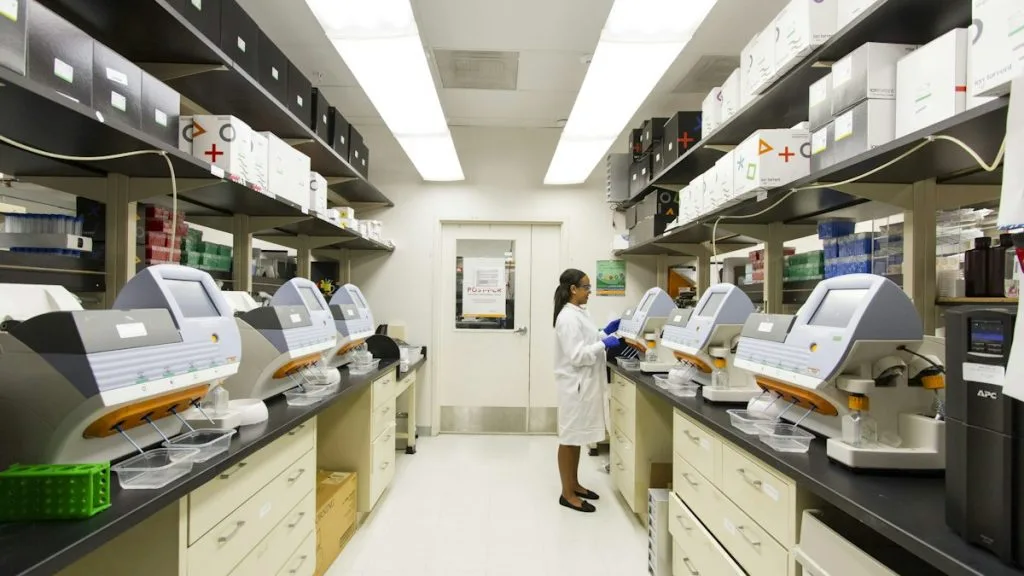
CLINICAL & LABORATORY DIAGNOSTIC PROTOCOL
- Specimen acquisition: Endoscopic mucosal biopsy or surgical resection specimen fixed in 10% neutral-buffered formalin (NBF) within 30 minutes of collection (cold ischemia time < 1 hr).
- Tissue processing: Formalin fixation time strictly calibrated to 24-48 hours at room temperature; paraffin embedding with 4-micron microtome sectioning on positively charged slides.
- Control standards: On-slide external positive control tonsil or placental tissue and validated negative isotype control run in parallel with every analytical batch.
Tissue Deparaffinization & Heat-Induced Epitope Retrieval (HIER)
Bake slides at 60°C for 60 minutes, deparaffinize through sequential xylene baths (3 x 5 min), and rehydrate through graded ethanols (100%, 95%, 70%) to deionized water. Perform HIER in target retrieval solution (pH 9.0 EDTA buffer) at 97°C for 20 minutes in a calibrated PT Link module.
Endogenous Peroxidase Block & Primary Antibody Incubation
Quench endogenous peroxidase activity using 3% hydrogen peroxide for 10 minutes. Apply optimized primary monoclonal antibody (**clone 22C3** or **clone SP263**) at calibrated clinical dilution and incubate for 30 minutes at room temperature in a humidified chamber.
Polymer Detection System & DAB Chromogen Visualization
Rinse in Tris-buffered saline with 0.05% Tween-20 (TBST). Apply horseradish peroxidase (HRP)-conjugated anti-mouse/rabbit polymer secondary reagent for 30 minutes. Incubate with freshly prepared 3,3-diaminobenzidine (DAB) substrate-chromogen solution for 10 minutes until brown precipitates develop.
Counterstaining, Dehydration & Quality Control Scoring
Counterstain nuclei with Mayer hematoxylin for 1-2 minutes, blue in running tap water, dehydrate through graded ethanols, clear in xylene, and coverslip with permanent non-aqueous mounting medium. Score tumor cell membrane and immune cell staining under light microscopy using the **Combined Positive Score (CPS)** algorithm.
1. Pathophysiology, Etiology & Cellular Carcinogenesis
Esophageal carcinogenesis involves distinct histopathologic pathways driven by chronic mucosal irritation, genomic instability, and accumulated somatic mutations. Squamous cell carcinomas arise from mucosal dysplasia, whereas adenocarcinomas predominantly evolve from metaplastic Barrett esophagus under chronic gastroesophageal reflux.
Epigenetic dysregulation and promoter hypermethylation frequently silence tumor suppressor loci such as **CDKN2A** and **FHIT**. Concurrent loss of cell cycle checkpoint regulation drives unchecked cellular proliferation and evasion of apoptosis.
The tumor microenvironment exhibits pronounced immunosuppressive features, characterized by transforming growth factor-beta (**TGF-beta**) secretion, regulatory T-cell infiltration, and myeloid-derived suppressor cell accumulation. Radiation therapy disrupts this stroma, exposing neoantigens but simultaneously inducing compensatory **PD-L1** upregulation.
2. Oncogenic Signal Transduction Pathways & Genomic Alterations
Receptor tyrosine kinase signaling networks, including **EGFR** and **HER2** (**ERBB2**), are frequently amplified or overexpressed in advanced esophageal malignancies. Ligand binding activates downstream intracellular cascades that promote survival, proliferation, and metabolic reprogramming.
The **PI3K-AKT-mTOR** and **RAS-RAF-MEK-ERK** pathways regulate protein synthesis, cellular growth, and resistance to radiation-induced apoptosis. Activating mutations within these axes correlate with aggressive clinical phenotypes and therapeutic resistance.
Genomic instability frequently manifests through **TP53** inactivation and chromosomal copy number alterations. Bypass signaling pathways often emerge post-chemoradiotherapy, necessitating combinatorial therapeutic approaches to prevent early relapse.
3. Histopathological Grading, Morphological Subtypes & Differential Diagnosis
Esophageal squamous cell carcinoma demonstrates intercellular bridges, keratinization, and nested architectural growth patterns. Adenocarcinoma exhibits glandular differentiation, mucin production, and complex cribriform or tubular structures.
Diagnostic immunohistochemical panels utilize markers such as **p40**, **CK5/6**, and **SOX2** to confirm squamous lineage. Adenocarcinoma diagnosis relies on positivity for **CK7**, **CDX2**, and **MUC5AC**.
Distinguishing primary esophageal lesions from metastatic carcinoma requires clinicopathologic correlation, endoscopic findings, and exact immunohistochemical expression profiling. Benign reactive atypia and radiation-induced cytologic changes must be carefully differentiated from residual invasive carcinoma.
4. Targeted Therapeutics, Landmark Phase III Trials & Resistance Mechanics
Immune checkpoint inhibitors such as **pembrolizumab** and **nivolumab** target the **PD-1/PD-L1** axis to reinvigorate exhausted tumor-specific T cells. These monoclonal antibodies block suppressive signals, permitting sustained anti-tumor immune responses following definitive chemoradiotherapy.
The landmark **CheckMate 577** Phase III trial established adjuvant **nivolumab** as standard of care after chemoradiotherapy and resection, demonstrating a superior median disease-free survival of 22.4 months versus 11.0 months for placebo. Similarly, the **KEYNOTE-590** trial validated frontline chemoimmunotherapy in advanced unresectable cohorts.
Adaptive resistance mechanisms include interferon-gamma receptor mutations, **beta-2-microglobulin** loss, and upregulation of alternative immune checkpoints such as **CTLA-4** or **LAG-3**. Combination strategies and novel bispecific antibodies are actively investigated to overcome these resistant phenotypes.
5. Molecular Staging, Minimal Residual Disease (ctDNA) & Prognostic Stratification
Precision oncology integrates AJCC 8th edition anatomic staging with molecular profiling to optimize prognostic stratification. Biomarkers such as **PD-L1 CPS**, **TMB**, and mismatch repair status guide adjuvant immunotherapy selection.
Liquid biopsy surveillance utilizing circulating tumor DNA (**ctDNA**) detects minimal residual disease and predicts recurrence prior to radiologic detection. Serial ctDNA quantification permits real-time assessment of therapeutic efficacy and early interception of progression.
Prognostic models incorporating clinical response to chemoradiotherapy, pathologic tumor regression grade, and genomic burden refine long-term survival expectations. Prospective clinical trials continue to evaluate risk-adapted duration of immunotherapy consolidation.