Collective-Amoeboid Transition in Invasive Carcinoma: Cellular Plasticity, Extracellular Matrix Remodeling & Molecular Determinants
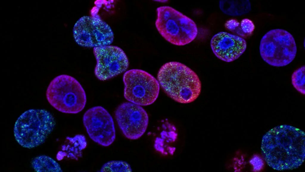
Collective-amoeboid transition (CAT) represents a dynamic cellular plasticity mechanism wherein carcinoma cells switch from cohesive, multicellular streaming migration to single-cell, mesenchymal-like blebbing locomotion. This phenotypic switch is governed by strict intracellular cytoskeletal rearrangement and extracellular matrix degradation.
- Primary molecular switch: Downregulation of epithelial cell-cell adhesion molecules including **CDH1** (**E-cadherin**) coupled with actin-myosin cytoskeleton contraction.
- Cytoskeletal regulation: Activation of the **RHOA**/**ROCK** signaling axis driving blebbing amoeboid movement versus **RAC1**/**CDC42** driving lamellipodial protrusion.
- Microenvironmental influence: Extracellular matrix pore size and density dictate the physical threshold required to trigger collective-to-amoeboid switching.
Carcinoma invasion relies on complex phenotypic plasticity that allows malignant cells to navigate diverse tissue architectures. The **collective-amoeboid transition** describes the capacity of tumor cell populations to alter their migration mode in response to mechanical and biochemical cues.
Mediated by alterations in cell adhesion and actomyosin contractility, this switch enables tumor cells to bypass dense extracellular matrix barriers. Understanding these signaling networks is essential for identifying actionable therapeutic targets to suppress metastatic dissemination.
CLINICAL & LABORATORY DIAGNOSTIC PROTOCOL
- Specimen acquisition: Fresh surgical resection or core needle biopsy fixed in 10% neutral-buffered formalin (**NBF**) within 15 minutes of devascularization to preserve labile phosphorylation states.
- Tissue processing: Formalin fixation optimized for 24–48 hours at room temperature, followed by paraffin embedding and microtome sectioning at 4-micron thickness onto superfrost plus glass slides.
- Control standards: Verified human tissue controls expressing high baseline levels of **E-cadherin** and **Vimentin** run concurrently with every immunohistochemical staining batch.
Deparaffinization & High-Temperature Epitope Unmasking
Bake slides at 60°C for 45 minutes, clear in xylene (3 changes, 5 minutes each), and rehydrate through descending ethanol concentrations (100%, 95%, 70%) to distilled water. Perform heat-induced epitope retrieval using pH 9.0 tris-EDTA buffer in a high-pressure decloaking chamber at 110°C for 15 minutes.
Endogenous Enzyme Blocking & Primary Antibody Application
Block endogenous peroxidase activity with 3% hydrogen peroxide for 10 minutes at room temperature, followed by washing in tris-buffered saline with Tween-20 (**TBST**). Apply validated primary monoclonal antibodies targeting **E-cadherin** (clone EP700Y) or **Vimentin** (clone V9) at standardized dilutions for 60 minutes.
Polymer Conjugate Incubation & Chromogenic Substrate Development
Apply horseradish peroxidase-conjugated goat anti-rabbit/mouse secondary polymer reagent for 30 minutes at room temperature. Rinse thoroughly with **TBST**, then apply freshly prepared 3,3-diaminobenzidine (**DAB**) chromogen substrate for 5 minutes to visualize antibody binding.
Nuclear Counterstaining, Dehydration & Mounting
Apply Mayer hematoxylin counterstain for 1 minute, rinse in running tap water for 5 minutes, dehydrate through ascending ethanols, clear in xylene, and apply resinous permanent mounting medium with a glass coverslip. Evaluate slides using brightfield microscopy for membrane versus cytoplasmic compartmentalization.
1. Pathophysiology, Etiology & Cellular Carcinogenesis
Carcinoma invasion involves structural remodeling of the surrounding stroma to facilitate migration away from the primary neoplastic site. Epithelial cancer cells frequently reside in cohesive cords or sheets before experiencing local microenvironmental triggers that induce single-cell motility.
Epigenetic silencing of tumor suppressor loci and upregulation of transcription factors such as **SNAI1** (**Snail**) and **ZEB1** drive the loss of intercellular junctions. This loss destabilizes cadherin-mediated cell-cell contacts and unleashes intrinsic migratory programs.
The tumor microenvironment provides mechanical and biochemical gradients, including hypoxia and elevated interstitial fluid pressure. These factors stimulate stromal fibroblasts and immune infiltrates to secrete matrix-degrading enzymes that accelerate the collective-amoeboid transition.
2. Oncogenic Signal Transduction Pathways & Genomic Alterations
Extracellular signaling ligands binding to receptor tyrosine kinases trigger intracellular cascades that orchestrate actin cytoskeletal dynamics. Phosphorylation events direct the activation status of small GTPases belonging to the Rho family.
The balance between **RHOA** and **RAC1** signaling dictates the prevailing mode of cellular locomotion. Upregulation of **ROCK1** (**Rho-associated protein kinase 1**) drives robust cortical actin contraction, characteristic of the rounded amoeboid phenotype.
Bypass signaling networks driven by **PI3K**/**AKT** and **MAPK** pathways sustain survival signals during matrix remodeling. These pathways prevent anoikis while cells undergo drastic mechanical deformation to squeeze through narrow extracellular matrix pores.
3. Histopathological Grading, Morphological Subtypes & Differential Diagnosis
Histopathological evaluation of invasive carcinomas reveals distinct architectural transition zones at the tumor-stroma interface. Pathologists identify streaming clusters breaking apart into isolated round or ellipsoid cells lacking intercellular bridges.
Immunohistochemical panels incorporating **CDH1**, **Vimentin**, and **MMP9** confirm the molecular shift from epithelial cohesion to mesenchymal or amoeboid plasticity. Loss of membranous **E-cadherin** concurrent with cytoplasmic **Vimentin** expression supports active transition.
Differential diagnosis requires distinguishing true collective-amoeboid invasion from sampling artifacts, crush injury, or stromal inflammation. Careful spatial mapping ensures accurate staging of invasive fronts and assessment of metastatic potential.
4. Targeted Therapeutics, Landmark Phase III Trials & Resistance Mechanics
Therapeutic strategies targeting cellular migration focus on inhibiting cytoskeletal regulators and matrix degradation enzymes. Small-molecule inhibitors directed against **ROCK** or matrix metalloproteinases aim to curtail invasive plasticity and metastatic seeding.
Landmark clinical investigations such as the **METEOR** and **LEAP** trials evaluated anti-angiogenic and multi-kinase inhibitors that indirectly normalize tumor vasculature and reduce hypoxia-driven migratory signals. Progression-free survival endpoints highlighted the challenge of overcoming adaptive metabolic and cytoskeletal pathways.
Acquired resistance to conventional cytotoxic chemotherapy often involves enrichment of subpopulations undergoing collective-amoeboid transition. Combination regimens incorporating targeted pathway inhibitors are currently under investigation to suppress escape mechanisms.
5. Molecular Staging, Minimal Residual Disease (ctDNA) & Prognostic Stratification
Integration of AJCC 8th edition TNM staging with molecular biomarkers of invasion provides superior risk stratification. Tumors exhibiting high frequencies of collective-amoeboid transition at the invasive front correlate with increased lymph node involvement and distant metastasis.
Liquid biopsy surveillance utilizing circulating tumor DNA (**ctDNA**) allows non-invasive tracking of genomic clones driving cellular plasticity and minimal residual disease. Serial quantification of mutated **RHOA** or **RAC1** allele frequencies offers real-time monitoring of metastatic progression.
Prognostic models incorporating tissue-based invasion patterns and liquid biomarker dynamics refine patient management paradigms. Prospective clinical validation continues to establish these cellular transition markers as core prognostic determinants in solid tumors.