Collective Cell Migration in Solid Tumors: Molecular Mechanics, E-Cadherin Dynamics & Therapeutic Implications
Collective cell migration defines the coordinated translocation of cohesive groups of cancer cells maintaining intercellular junctions. This invasive mechanism drives aggressive metastasis across epithelial malignancies.
- Primary cellular mechanism: Retention of cell-cell contacts via intact E-cadherin junctional complexes during tissue invasion.
- Cytoskeletal regulation: Coordinated activation of Rho-family GTPases (RhoA, Rac1, Cdc42) driving leader-follower phenotypes.
- Therapeutic implication: Resistance to single-agent mesenchymal transition inhibitors due to phenotypic plasticity and cooperative survival.
Collective cell migration represents a predominant mode of cancer invasion where multicellular strands, sheets, or clusters invade adjacent stroma. This multicellular movement contrasts with single-cell mesenchymal or amoeboid migration by preserving intercellular adhesion structures.
The process relies on continuous biochemical cross-talk between leader cells at the invasion front and follower cells within the bulk. Spatial coordination directs extracellular matrix remodeling, mechanical force generation, and directional persistence through tissue microenvironments.
CLINICAL & LABORATORY DIAGNOSTIC PROTOCOL
- Specimen acquisition: Fresh surgical resection or diagnostic core biopsy fixed in 10% neutral-buffered formalin (NBF) with a cold ischemia time strictly maintained under 45 minutes.
- Tissue processing: Automated dehydration, clearing, and paraffin infiltration protocols yielding 4-micron thick sections on silane-coated adhesive slides.
- Control standards: Internal normal epithelial tissue controls and validated external positive controls included in every immunohistochemical run.
Slide Preparation and Antigen Retrieval
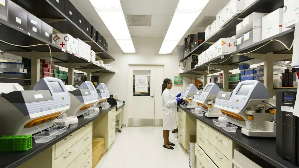
Bake histological sections at 60°C for 45 minutes, pass through xylene (3 changes, 3 minutes each), and rehydrate through graded ethanols (100%, 95%, 70%) to distilled water. Perform heat-induced epitope retrieval in a high-pH 9.0 Tris-EDTA buffer at 97°C for 25 minutes using a pressure-controlled laboratory instrument.
Blocking and Primary Antibody Incubation
Block endogenous peroxidase activity with 3.0% hydrogen peroxide for 10 minutes at room temperature, followed by rinsing in phosphate-buffered saline with 0.1% Tween-20 (PBST). Incubate sections with primary monoclonal antibodies against **E-cadherin** (clone NCH-38) or **Vimentin** (clone V9) at optimized dilutions for 60 minutes.
Polymer Detection and Chromogen Development
Apply horseradish peroxidase (HRP)-conjugated goat anti-mouse/rabbit secondary polymer for 30 minutes at room temperature. Wash thoroughly in PBST, then apply freshly prepared 3,3-diaminobenzidine (DAB) chromogen solution for exactly 5 minutes to visualize antibody binding.
Counterstaining and Evaluation
Counterstain nuclei with modified Lillie-Mayer hematoxylin for 60 seconds, blue in alkaline tap water, dehydrate through graded alcohols, clear in xylene, and apply resinous permanent coverslip. Evaluate collective migration fronts under light microscopy, recording membranous retention versus loss of adhesion molecules.
1. Pathophysiology, Etiology & Cellular Carcinogenesis
Collective cell migration emerges from genetic and epigenetic alterations that subvert normal epithelial homeostasis. Tumor cells retain functional intercellular adhesions while acquiring polarity, allowing them to invade as cohesive multicellular units.
Epigenetic dysregulation frequently modulates cell-surface receptors, balancing adhesive forces with mechanical traction against the extracellular matrix. Promoter hypermethylation of tumor suppressor loci further stabilizes this invasive phenotype.
The surrounding tumor microenvironment provides biochemical gradients of chemokines and growth factors that polarize the migrating cohort. Cancer-associated fibroblasts remodel collagen tracks, facilitating physical movement of the leading edge.
2. Oncogenic Signal Transduction Pathways & Genomic Alterations
Upstream receptor tyrosine kinase activation triggers complex downstream signaling networks that govern cytoskeletal dynamics. Signaling fluxes through the **PI3K-AKT-mTOR** and **RAS-RAF-MEK-ERK** pathways dictate mechanical force generation and directional migration.
Intracellular coordination is heavily mediated by **Rho-family GTPases**, where **RhoA** controls actomyosin contractility at the rear, while **Rac1** and **Cdc42** drive lamellipodia and filopodia extension at the front.
Mutations in regulatory genes or loss of core adhesion proteins like **CDH1** modulate the transition between single and collective migration modes, enhancing metastatic efficiency across various organ sites.
3. Histopathological Grading, Morphological Subtypes & Differential Diagnosis
Histological evaluation of collective migration reveals streaming cords, solid nests, or glandular strands penetrating stromal barriers. Architectural patterns often display distinct leading-edge polarization distinct from the central tumor mass.
Immunohistochemical panels utilizing **E-cadherin**, **N-cadherin**, and **cytokeratins** confirm epithelial preservation within migrating clusters. Absence of mesenchymal markers helps differentiate collective strands from true sarcomatoid dedifferentiation.
Differential diagnosis requires distinguishing invasive strands from entrapped benign ducts, reactive mesothelial hyperplasia, and vascular pseudo-invasion artifacts. Precise anatomical localization and cytological atypia assessment ensure accurate diagnostic grading.
4. Targeted Therapeutics, Landmark Phase III Trials & Resistance Mechanics
Therapeutic targeting of collective cell migration focuses on disrupting actomyosin contractility and cell-matrix interactions. Pharmacological inhibition of **ROCK (Rho-associated protein kinase)** and targeted multi-kinase inhibitors impair leading-edge force generation.
Landmark Phase III trials evaluating anti-stromal agents and metastasis inhibitors have demonstrated complex dynamics regarding progression-free survival in aggressive solid tumors. Clinical outcomes emphasize the redundancy of migratory signaling networks.
Resistance to anti-migratory therapies frequently arises through phenotypic plasticity, allowing tumors to switch seamlessly between collective and single-cell amoeboid migration modes under therapeutic pressure.
5. Molecular Staging, Minimal Residual Disease (ctDNA) & Prognostic Stratification
Integration of collective migration markers with **AJCC 8th Edition** TNM staging refines prognostic stratification for high-risk patients. Presence of extensive collective invasion correlates with increased incidence of regional lymph node metastasis.
Liquid biopsy surveillance utilizing **circulating tumor DNA (ctDNA)** tracks clonal evolution and detects minimal residual disease before radiological recurrence. High-throughput sequencing identifies mutations driving invasive phenotypes.
Prospective clinical paradigms increasingly incorporate spatial transcriptomics and deep tissue pathology to map invasive fronts. These diagnostic modalities guide personalized therapeutic regimens aimed at suppressing metastatic dissemination.