Colorectal Carcinoma: Microsatellite Instability (MSI-High), Mismatch Repair Deficiency & Nivolumab
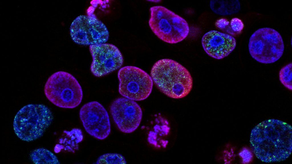
Colorectal carcinoma characterized by high microsatellite instability (MSI-H) and mismatch repair deficiency (dMMR) accounts for approximately 15 percent of all colorectal malignancies. These tumors exhibit hypermutator phenotypes responsive to immune checkpoint inhibition using anti-PD-1 monoclonal antibodies.
- Molecular hallmark: Inactivation of DNA mismatch repair genes leading to microsatellite instability (MSI-H).
- Frontline immunotherapy: Anti-PD-1 monoclonal antibody nivolumab monotherapy or combined with ipilimumab.
- Prognostic profile: Stage-dependent survival advantage with immunotherapy over conventional cytotoxic chemotherapy.
Microsatellite instability-high (MSI-H) colorectal carcinoma arises from germline mutations in mismatch repair genes (Lynch syndrome) or somatic hypermethylation of the MLH1 promoter. Demographic distributions span hereditary cohorts presenting at younger ages and sporadic cases clustering among older females.
Defective DNA mismatch repair causes length alterations within repetitive DNA sequences termed microsatellites across the genome. The resulting accumulation of frameshift mutations generates neoantigens that stimulate robust tumor-infiltrating lymphocyte activation.
CLINICAL & LABORATORY DIAGNOSTIC PROTOCOL
- Specimen acquisition: Formalin-fixed paraffin-embedded (FFPE) tumor tissue blocks or unstained serial sections derived from surgical resection or core biopsy specimens.
- Fixation parameters: Tissue immersion in 10% neutral-buffered formalin for a mandatory fixation duration of 6 to 72 hours under controlled ambient temperature.
- Reagent validation: Positive control tissue containing normal colonic mucosa with intact nuclear expression for MLH1, MSH2, MSH6, and PMS2 run alongside analytical batches.
Deparaffinization and Heat-Induced Epitope Retrieval
Bake FFPE sections at 60°C for 45 minutes, pass through three changes of xylene for 5 minutes each, and rehydrate through descending ethanol dilutions to distilled water. Perform heat-induced epitope retrieval in a high-pH EDTA buffer (pH 9.0) at 97°C for 25 minutes using an automated slide stainer.
Peroxide Blockade and Primary Antibody Application
Block endogenous peroxidase activity by incubating slides in 3% hydrogen peroxide for 10 minutes at room temperature, followed by washing in Tris-buffered saline with Tween-20. Apply optimized monoclonal primary antibodies directed against MLH1 (clone ES05), MSH2 (clone FE11), MSH6 (clone EP49), and PMS2 (clone EP51) for 30 minutes.
Polymer Detection and Chromogenic Visualization
Incubate sections with a horseradish peroxidase-conjugated secondary polymer reagent for 30 minutes at room temperature. Visualize antibody binding by applying 3,3-diaminobenzidine substrate-chromogen solution for 10 minutes to yield a distinct brown nuclear precipitate.
Nuclear Counterstaining and Pathologist Evaluation
Apply Mayer hematoxylin nuclear counterstain for 1 minute, dehydrate through graded ethanols, clear in xylene, and mount with a resinous medium. Evaluate nuclear staining in viable invasive carcinoma cells using internal stromal lymphocytes and normal epithelial crypts as positive internal controls.
1. Pathophysiology, Etiology & Cellular Carcinogenesis
Defective mismatch repair disrupts the cellular surveillance network responsible for correcting single-nucleotide mismatches and insertion-deletion loops during DNA replication. Somatic inactivation typically stems from hypermethylation of the MLH1 promoter region accompanied by acquired BRAF V600E mutations in sporadic tumors. Lynch syndrome involves autosomal dominant germline pathogenic variants in MLH1, MSH2, MSH6, or PMS2.
Epigenetic dysregulation silences tumor suppressor loci and accelerates transition through classic adenoma-carcinoma and serrated neoplasia pathways. Loss of DNA proofreading capacity drives widespread frameshift mutations in coding microsatellites of genes regulating cell growth and apoptosis. The resulting genomic instability fosters rapid subclonal evolution and malignant transformation.
The tumor microenvironment in mismatch repair-deficient colorectal carcinoma features dense infiltration by cytotoxic CD8-positive T lymphocytes and Th1 helper cells. Upregulation of immune checkpoints including PD-1 and PD-L1 creates an adaptive immune resistance network. This inflamed stromal architecture establishes the biological rationale for therapeutic blockade using immune checkpoint inhibitors.
2. Oncogenic Signal Transduction Pathways & Genomic Alterations
Genomic hypermutation in mismatch repair-deficient tumors produces high densities of neoepitopes that engage host immune receptors. Concurrently, activating mutations in growth factor receptor pathways such as PI3K-AKT-mTOR and RAS-RAF-MEK-ERK maintain constitutive proliferative signaling. Secondary alterations often involve TGF-beta receptor type II and IGFR signaling domains mutated at coding microsatellite tracts.
Intracellular signal transduction cascades integrate environmental cues to regulate gene transcription, cell cycle progression, and metabolic reprogramming. Unchecked activation of downstream kinase cascades promotes survival against apoptotic stimuli and enhances invasive cellular motility. Crosstalk between tumor cells and cancer-associated fibroblasts further reinforces extracellular matrix remodeling.
Resistance mechanisms to immunotherapy emerge through acquired beta-2-microglobulin mutations that abrogate major histocompatibility complex class I antigen presentation. Loss of interferon-gamma signaling pathway components also contributes to acquired therapeutic resistance. Identifying these secondary genomic events directs salvage interventions in refractory disease.
3. Histopathological Grading, Morphological Subtypes & Differential Diagnosis
Histopathological examination frequently reveals high-grade mucinous, signet-ring, or medullary growth patterns characterized by Crohn-like lymphocytic reactions. Tumor-infiltrating lymphocytes are abundant at the invasive front and within intratumoral compartments. Cytological features include large pleomorphic nuclei, prominent nucleoli, and abundant eosinophilic cytoplasm.
Diagnostic evaluation utilizes immunohistochemistry evaluating MLH1, MSH2, MSH6, and PMS2 protein expression alongside secondary PCR or next-generation sequencing assays. Correlation with reflex BRAF V600E mutation testing distinguishes sporadic hypermethylation from Lynch syndrome. Exclusion of neuroendocrine differentiation and squamous metaplasia ensures precise histological classification.
Differential diagnoses include poorly differentiated neuroendocrine carcinomas, metastatic adenocarcinomas from extraintestinal primaries, and primary colonic lymphomas. Comprehensive immunohistochemical panels incorporating cytokeratins CK7 and CK20, CDX2, and SATB2 confirm colorectal lineage. Accurate histological categorization directly dictates systemic treatment algorithms.
4. Targeted Therapeutics, Landmark Phase III Trials & Resistance Mechanics
Frontline therapeutic intervention for metastatic mismatch repair-deficient colorectal carcinoma relies on anti-PD-1 monoclonal antibodies such as nivolumab. Nivolumab blocks inhibitory signaling interactions between PD-1 on activated T cells and PD-L1 ligands on tumor cells. Combination regimens incorporating ipilimumab provide dual immune checkpoint blockade via simultaneous CTLA-4 inhibition.
The landmark CheckMate-142 clinical trial demonstrated deep, durable objective response rates and prolonged progression-free survival with nivolumab combination regimens. Subsequent Phase III data confirmed superior overall survival and manageable toxicity profiles compared to traditional cytotoxic chemotherapy backbones. These findings established anti-PD-1 immunotherapy as the global standard of care.
Primary resistance occurs in a small subset of mismatch repair-deficient tumors possessing alternative oncogenic driver mutations or primary immune evasion phenotypes. Secondary resistance involves structural alterations in antigen-presenting machinery and loss of interferon pathway signaling. Ongoing clinical investigations evaluate novel bispecific antibodies and targeted combination therapies to overcome acquired resistance.
5. Molecular Staging, Minimal Residual Disease (ctDNA) & Prognostic Stratification
Staging integration combines AJCC 8th edition anatomic parameters with mismatch repair status and tumor mutational burden metrics. Mismatch repair deficiency confers a favorable prognosis in localized stages II and III disease compared to proficient counterparts. Precise molecular stratification guides decisions regarding adjuvant chemotherapy administration versus active surveillance.
Liquid biopsy surveillance utilizing circulating tumor DNA enables sensitive detection of minimal residual disease following surgical resection. Serial tracking of tumor-specific somatic variants provides early lead-time detection of clinical recurrence prior to radiographic visualization. Quantitative ctDNA kinetics also serve as surrogate biomarkers for monitoring immunotherapy efficacy.
Prognostic models incorporate clinicopathological variables, microsatellite stability status, and circulating biomarker dynamics into predictive risk calculators. Prospective validation of these integrated algorithms refines postoperative management strategies and personalizes surveillance schedules. Continued clinical trial enrollment remains essential for optimizing long-term survival outcomes.