Contact Inhibition Breakdown and Cell-Density Regulation in Epithelial Carcinogenesis
Contact normalization loss defines malignant epithelial transformation, whereby loss of density-dependent growth arrest drives uncontrolled proliferation. This biophysical and biochemical breakdown precipitates dysregulated tissue homeostasis.
- Loss of E-cadherin-mediated intercellular adhesion triggers the nuclear translocation of YAP1 and TAZ transcription co-activators.
- Mechanical stretch and extracellular matrix rigidity override standard cellular crowding signals, maintaining high mitotic indices.
- Targeting hippo signaling effectors provides novel therapeutic avenues in treatment-refractory solid tumors.
Contact inhibition failure represents a cardinal hallmark of oncogenic transformation in diverse epithelial tissues. Histopathological analysis consistently demonstrates a loss of monolayer architectural restraint, leading to multilayered, invasive cellular cords.
The disruption of homotypic cell-cell contacts overrides normal mechanical feedback loops. This biophysical failure permits sustained signal transduction cascades despite extreme cellular density.
CLINICAL & LABORATORY DIAGNOSTIC PROTOCOL
- Specimen acquisition: Core needle biopsy or surgical resection fixed in 10% neutral-buffered formalin (NBF) within 30 minutes of devascularization (cold ischemia time < 1 hr).
- Tissue processing: Formalin fixation time strictly calibrated to 24–48 hours at room temperature; paraffin embedding with 4-micron microtome sectioning on positively charged slides.
- Control standards: On-slide external positive control tissue and validated negative isotype control run in parallel with every analytical batch.
Tissue Deparaffinization & Heat-Induced Epitope Retrieval (HIER)
Bake slides at 60°C for 60 minutes, deparaffinize through sequential xylene baths (3 x 5 min), and rehydrate through graded ethanols (100%, 95%, 70%) to deionized water. Perform HIER in target retrieval solution (pH 9.0 EDTA buffer or pH 6.0 citrate buffer) at 97°C for 20 minutes in a calibrated PT Link module.
Endogenous Peroxidase Block & Primary Antibody Incubation
Quench endogenous peroxidase activity using 3% hydrogen peroxide for 10 minutes. Apply optimized primary monoclonal antibody (e.g., anti-E-cadherin clone NCH-38, or anti-YAP1 clone D8H1X) at calibrated clinical dilution and incubate for 30–60 minutes at room temperature in a humidified chamber.
Polymer Detection System & DAB Chromogen Visualization
Rinse in Tris-buffered saline with 0.05% Tween-20 (TBST). Apply horseradish peroxidase (HRP)-conjugated anti-rabbit/mouse polymer secondary reagent for 30 minutes. Incubate with freshly prepared 3,3-diaminobenzidine (DAB) substrate-chromogen solution for 5-10 minutes until brown precipitates develop.
Counterstaining, Dehydration & Quality Control Scoring
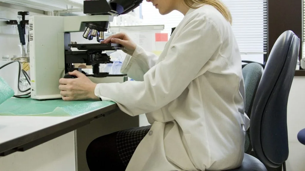
Counterstain nuclei with Mayer hematoxylin for 1–2 minutes, blue in running tap water or 0.08% ammonia water, dehydrate through graded ethanols, clear in xylene, and coverslip with permanent non-aqueous mounting medium. Score tumor cell membrane/nuclear staining under light microscopy using validated scoring algorithm.
1. Pathophysiology, Etiology & Cellular Carcinogenesis
Epithelial tissues rely on intercellular adhesion complexes to maintain architectural integrity and restrict mitotic output upon reaching confluence. Mutational inactivation of the **CDH1** gene encoding **E-cadherin** abolishes calcium-dependent cellular binding. This structural failure removes the primary mechanical trigger required for contact-mediated growth arrest.
Epigenetic silencing via promoter hypermethylation of tumor suppressor loci reinforces this malignant phenotype. Concurrent downregulation of cell cycle inhibitors such as **p27** prevents density-dependent cell cycle exit at the G1/S boundary.
The surrounding tumor microenvironment actively promotes this breakdown through cancer-associated fibroblasts and stiffened collagen matrices. Extracellular matrix remodeling overrides normal biophysical density sensing mechanisms, perpetuating aggressive clonal expansion.
2. Oncogenic Signal Transduction Pathways & Genomic Alterations
Normal intercellular contact activates the **Hippo pathway**, resulting in the phosphorylation and cytoplasmic retention of **YAP1** and **TAZ**. In contact-inhibited cells, kinase cascades involving **LATS1/2** prevent these transcriptional co-activators from translocating to the nucleus. Carcinogenic mutations bypass this negative regulation entirely.
When cell-cell contacts fail, unphosphorylated **YAP1** enters the nucleus to bind **TEAD** transcription factors. This complex drives the transcription of pro-proliferative and anti-apoptotic genes, including **CCND1** and **BCL2**.
Secondary bypass signaling through **PI3K-AKT-mTOR** and **RAS-MAPK** axes synergizes with nuclear YAP1 accumulation. These concurrent alterations render epithelial cancer cells completely refractory to spatial crowding constraints.
3. Histopathological Grading, Morphological Subtypes & Differential Diagnosis
Histological examination of contact-inhibition-deficient carcinomas reveals disorganized architectural growth patterns, including solid, micropapillary, and cribriform configurations. Marked cellular pleomorphism, high nuclear-to-cytoplasmic ratios, and atypical mitotic figures are diagnostic hallmarks. Invasive fronts demonstrate loss of distinct basement membrane demarcation.
Diagnostic immunohistochemical panels utilize **TTF-1**, **Napsin A**, and **CK7** for pulmonary lesions, alongside **E-cadherin** loss to confirm epithelial-to-mesenchymal transition. Aberrant cytoplasmic or absent membranous **E-cadherin** staining confirms the structural breakdown of intercellular junctions. Proliferative fraction assessment via **Ki-67** routinely exceeds 30 percent in high-grade lesions.
Differential diagnosis requires distinguishing primary poorly differentiated carcinomas from metastatic deposits and reactive mesothelial proliferations. Specialized markers such as **WT1** and **Calretinin** help exclude mesothelial mimics, while molecular profiling confirms primary tissue origin.
4. Targeted Therapeutics, Landmark Phase III Trials & Resistance Mechanics
Directly targeting the transcriptional outputs of failed contact inhibition remains an active area of clinical investigation. Small molecule inhibitors targeting **FAK** and upstream **mTOR** pathways attenuate aberrant mechanotransduction signaling. Combination regimens involving **trametinib** and **everolimus** aim to block downstream survival cascades.
Landmark Phase III trials such as **KEYNOTE-189** and **CheckMate-9LA** highlight the efficacy of combining platinum doublet chemotherapy with immune checkpoint inhibitors in tumors lacking contact inhibition. Progression-free survival and overall survival metrics demonstrate marked improvement over chemotherapy alone in PD-L1 positive cohorts. However, acquired resistance frequently emerges via alternative receptor tyrosine kinase upregulation.
Tertiary resistance often develops through secondary mutations in downstream kinases or epithelial-to-mesenchymal transition loops. Novel antibody-drug conjugates directed against surface antigens bypass traditional intracellular resistance mechanisms to restore tumor control.
5. Molecular Staging, Minimal Residual Disease (ctDNA) & Prognostic Stratification
Integration of **AJCC 8th edition** TNM staging with molecular biomarkers provides precise prognostic stratification for aggressive carcinomas. Pathological evaluation of resection margins and lymph node status remains mandatory for establishing recurrence risk. High nuclear grade and diffuse loss of adhesion molecules correlate with poor clinical outcomes.
Liquid biopsy surveillance utilizing circulating tumor DNA (**ctDNA**) allows for real-time detection of minimal residual disease following surgical resection. Next-generation sequencing panels track tumor-specific mutations to identify molecular relapse prior to radiographic progression. Serial ctDNA monitoring guides therapeutic adjustments in real time.
Overall survival prognostic algorithms increasingly incorporate multi-omic profiles to personalize postoperative treatment strategies. Prospective clinical validation of these tissue-based and circulating biomarkers continues to refine oncology clinical paradigms.