Definitive Carbon Ion Radiotherapy for Thoracic Bone and Soft Tissue Sarcoma: Radiobiology, Biophysical Mechanics & Clinical Outcomes
Definitive carbon ion radiotherapy utilizes high linear energy transfer (LET) particles to overcome radioresistance in complex thoracic bone and soft tissue sarcomas. This advanced modality delivers precise Bragg peak dose deposition, achieving high local control rates while sparing critical mediastinal structures.
- Radiobiological advantage: High relative biological effectiveness (RBE) driven by dense ionization clusters causing complex, irreparable double-strand DNA breaks.
- Geometric precision: Sharp lateral penumbra and inverse depth-dose profile of carbon ions protect adjacent spinal cord, heart, and great vessels.
- Clinical efficacy: Superior local tumor control in radioresistant histologies such as chondrosarcoma, osteosarcoma, and retroperitoneal or thoracic wall dedifferentiated liposarcoma.
Thoracic sarcomas comprise a rare, heterogeneous group of malignant mesenchymal neoplasms arising from the chest wall, ribs, vertebrae, and intrathoracic soft tissues. Conventional photon and proton therapies often fail to deliver adequate ablative doses due to strict radiation dose constraints of adjacent critical cardiopulmonary structures.
Carbon ion radiotherapy overcomes these limitations through high-LET radiation physics, exhibiting a dense ionization track that induces complex DNA lesions independent of cellular oxygenation status. This biological advantage minimizes the oxygen enhancement ratio (OER) disadvantage seen in hypoxic tumor microenvironments characteristic of large sarcomas.
CLINICAL & LABORATORY DIAGNOSTIC PROTOCOL
- Specimen acquisition: Core needle biopsy or wide surgical resection fixed in 10% neutral-buffered formalin (NBF) within 20 minutes of collection to prevent autolysis.
- Tissue processing: Formalin fixation maintained between 24 and 48 hours, followed by paraffin embedding and microtome sectioning at 4-micron thickness on charged glass slides.
- Control standards: Internal vimentin or desmin positive controls and non-immune serum negative controls run simultaneously for immunohistochemical verification.
Deparaffinization and Heat-Induced Epitope Retrieval (HIER)
Bake slides at 60°C for 45 minutes, pass through three changes of xylene for 5 minutes each, and rehydrate through graded ethanol solutions (100%, 95%, 80%) into distilled water. Perform HIER in a 99°C PT module using pH 9.0 Tris-EDTA buffer for 25 minutes.
Peroxidase Blocking and Primary Antibody Application
Block endogenous peroxidase activity by incubating sections in 3.0% hydrogen peroxide solution for 10 minutes at room temperature. Apply optimized primary monoclonal antibodies against sarcoma-specific markers (e.g., **INI1** clone MRQ-27, **MDM2** clone IF2) at predetermined dilutions.
Polymer Detection and Chromogenic Substrate Incubation
Wash slides thoroughly in phosphate-buffered saline with 0.05% Tween-20 (PBST). Apply horseradish peroxidase (HRP)-conjugated goat anti-rabbit/mouse polymer secondary reagent for 30 minutes, followed by exposure to 3,3-diaminobenzidine (DAB) chromogen substrate for 7 minutes.
Nuclear Counterstaining and Permanent Coverslipping
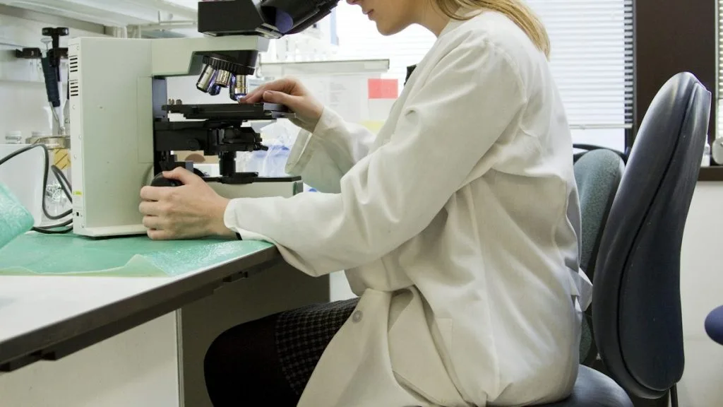
Counterstain nuclear structures with Mayer hematoxylin for 90 seconds, blue in warm running tap water, dehydrate through ascending ethanols and xylene, and mount with resinous mounting media. Evaluate staining intensity and cellular distribution using light microscopy.
1. Pathophysiology, Etiology & Cellular Carcinogenesis
Mesenchymal transformation in thoracic sarcomas involves complex chromosomal translocations, gene amplifications, or loss-of-function mutations driving uncontrolled cellular proliferation. Structural rearrangements such as the **SS18-SSX** fusion gene dictate synovial sarcoma oncogenesis via epigenetic remodeling and aberrant transcriptional activation.
Genomic instability frequently manifests as chromothripsis or complex karyotypes, particularly in pleomorphic and dedifferentiated soft tissue subtypes. Epigenetic silencing of tumor suppressor loci through DNA methyltransferase activity further accelerates malignant progression within the thoracic wall and mediastinal compartments.
The tumor microenvironment features dense, hypovascular stroma characterized by cancer-associated fibroblasts and profound hypoxia. This hypoxic niche upregulates **HIF-1alpha**, driving angiogenesis and fostering an immunosuppressive microenvironment resistant to conventional therapies.
2. Oncogenic Signal Transduction Pathways & Genomic Alterations
Aberrant activation of receptor tyrosine kinases and downstream signaling networks sustains survival and proliferation in thoracic sarcomas. The **PI3K-AKT-mTOR** pathway is frequently hyperactivated through genomic gains or PTEN loss, promoting unchecked cell growth and evasion of apoptosis.
Concurrently, the **RAS-RAF-MEK-ERK** cascade stimulates transcriptional machinery required for cell cycle progression and extracellular matrix remodeling. Cyclin-dependent kinase dysregulation, notably **CDK4** amplification paired with **p16** loss, drives accelerated G1-S phase transition.
Impaired DNA damage response mechanisms, stemming from structural **TP53** mutations or **MDM2** overexpression, prevent cell cycle arrest and apoptosis following genomic insult. Carbon ion radiotherapy directly overrides these survival pathways by inflicting complex, clustered DNA breaks that bypass standard repair kinetics.
3. Histopathological Grading, Morphological Subtypes & Differential Diagnosis
Thoracic sarcomas encompass distinct morphological subtypes including chondrosarcoma, osteosarcoma, malignant peripheral nerve sheath tumor, and synovial sarcoma. Architectural evaluation reveals fascicular, spindle cell, or epithelioid growth patterns coupled with high nuclear pleomorphism and frequent atypical mitotic figures.
Histopathological grading relies on the Fédération Nationale des Centres de Lutte contre le Cancer (FNCLCC) system, integrating differentiation score, mitotic count, and tumor necrosis extent. High-grade tumors (Grade 3) exhibit extensive necrosis and greater than 20 mitoses per 10 high-power fields.
Diagnostic immunohistochemical panels are mandatory to differentiate sarcomas from poorly differentiated carcinomas and lymphomas. Specific markers such as **SATB2** for osteoblastic differentiation and **INI1** retention status guide definitive pathological classification.
4. Targeted Therapeutics, Landmark Phase III Trials & Resistance Mechanics
Systemic management of advanced thoracic sarcomas traditionally involves anthracycline-based chemotherapy regimens including Doxorubicin and Ifosfamide. Multi-agent clinical trials establish these cytotoxic agents as frontline standards for metastatic disease, though local recurrence remains the primary pattern of failure.
Landmark Phase III trials evaluating precision tyrosine kinase inhibitors, such as Pazopanib, demonstrate significant progression-free survival benefits in non-adipocytic soft tissue sarcomas. Combination strategies incorporating immune checkpoint inhibitors targeting the **PD-1/PD-L1 axis** continue to be investigated in clinical cohorts.
Intrinsic radioresistance to photon irradiation is mediated by rapid sublethal damage repair and robust antioxidant defense mechanisms within sarcoma stem cell niches. Carbon ion radiotherapy circumvents this resistance by depositing high-density energy directly along the particle trajectory, inducing cell death independently of repair capacity.
5. Molecular Staging, Minimal Residual Disease (ctDNA) & Prognostic Stratification
Staging algorithms integrate anatomical tumor size, depth, nodal involvement, and distant metastasis with molecular profiling metrics. The **AJCC 8th edition** criteria categorize tumors based on histological grade to establish accurate prognostic groupings and therapeutic strategies.
Liquid biopsy surveillance utilizing circulating tumor DNA (**ctDNA**) analysis permits sensitive detection of minimal residual disease following definitive radiation therapy. Quantification of tumor-specific genomic breakpoints enables early identification of recurrence before radiographic visualization.
Multidisciplinary prognostic stratification models combine genomic aberrations, pathological grade, and dosimetric parameters from carbon ion treatment plans. These integrated scores optimize patient selection for definitive particle therapy and post-treatment surveillance paradigms.