EDNRA Forms a Positive Feedback Loop with the Hippo/YAP Axis to Drive Triple-Negative Breast Cancer Progression
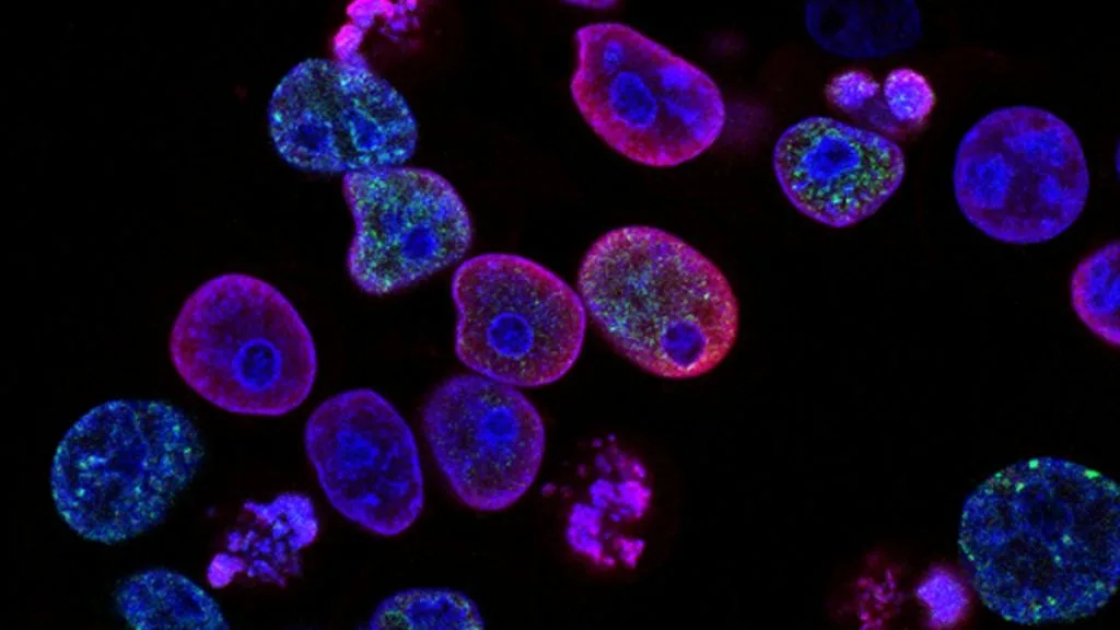
Triple-negative breast cancer remains a formidable clinical challenge driven by aggressive stromal remodeling and transcriptional co-activator hyperactivation. Aberrant **EDNRA** (endothelin receptor type A) overexpression engages a feed-forward signaling loop with the **Hippo/YAP** (Yes-associated protein) transcriptional axis to accelerate tumor progression.
- Molecular axis: **EDNRA** activation translocates **YAP** and **TAZ** into the nucleus, binding **TEAD** transcription factors to upregulate pro-metastatic target genes.
- First-line chemo-immunotherapy: **pembrolizumab** combined with platinum agents improves pathological complete response in PD-L1 positive tumors.
- Antibody-drug conjugates: **sacituzumab govitecan** targets **Trop-2** to deliver SN-38 directly to chemotherapy-resistant malignant cells.
Triple-negative breast cancer represents approximately 15 percent of all invasive breast malignancies and disproportionately affects premenopausal women under the age of 40. Patients lacking estrogen receptor, progesterone receptor, and HER2 expression exhibit heightened rates of visceral metastasis and early recurrence.
Oncogenic drivers within the microenvironment frequently hijack developmental signaling networks to sustain cellular proliferation. The G-protein coupled receptor **EDNRA** aberrantly interfaces with mechanotransduction cascades to bypass conventional growth inhibitor checkpoints.
CLINICAL & LABORATORY DIAGNOSTIC PROTOCOL
- Specimen acquisition: Core needle biopsy or radical resection tissue fixed in 10% neutral-buffered formalin (NBF) within 15 minutes of devascularization to preserve **EDNRA** and **YAP** nuclear localization.
- Tissue processing: Formalin fixation time rigorously controlled between 24 and 48 hours at 22°C, followed by paraffin embedding and microtome sectioning at 4-micron thickness onto superfrost plus slides.
- Control standards: Human leiomyosarcoma tissue used as a verified positive control for **EDNRA**, and tonsil tissue for immune cell markers, run alongside every diagnostic run.
Deparaffinization and Heat-Induced Epitope Retrieval (HIER)
Bake slides at 60°C for 45 minutes, pass through three changes of xylene for 5 minutes each, and rehydrate through graded absolute to 70 percent ethanol solutions. Perform HIER in high-pH Tris-EDTA buffer (pH 9.0) at 98°C for 25 minutes using an automated pressure cooker module.
Endogenous Block and Primary Antibody Incubation
Block endogenous peroxidase activity with 3 percent hydrogen peroxide in methanol for 10 minutes at room temperature. Apply anti-**EDNRA** rabbit monoclonal primary antibody (clone EPR13958) or anti-**YAP** (clone D8H1X) at 1:200 dilution for 60 minutes in a humidified chamber.
Polymer Detection and Chromogen Development
Wash slides thoroughly in Tris-buffered saline with 0.05 percent Tween-20 (TBST). Apply horseradish peroxidase-conjugated anti-rabbit secondary polymer for 30 minutes, followed by incubation with 3,3-diaminobenzidine (DAB) chromogen substrate for exactly 6 minutes.
Counterstaining and Pathologist Evaluation
Counterstain nuclear structures with modified Mayer hematoxylin for 90 seconds, wash in alkaline bluing reagent, dehydrate through ethanols, clear in xylene, and mount with synthetic resin. Evaluate percentage of positive invasive tumor cells and subcellular localization.
1. Pathophysiology, Etiology & Cellular Carcinogenesis
Triple-negative breast cancer pathogenesis involves extensive genomic instability driven by frequent **TP53** mutations and homologous recombination deficiencies. Basal-like transcriptomic subtypes demonstrate high mitotic indices, prominent central necrosis, and pushing tumor borders.
Epigenetic silencing of tumor suppressor loci through promoter hypermethylation cooperates with aberrant activation of embryonic pathways. The resulting cellular plasticity permits rapid adaptation to hypoxic microenvironments within the advancing tumor front.
The tumor stroma is heavily infiltrated by immunosuppressive tumor-associated macrophages and cancer-associated fibroblasts. These stromal elements secrete high levels of endothelin-1, which directly stimulates epithelial **EDNRA** expression on malignant ductal cells.
2. Oncogenic Signal Transduction Pathways & Genomic Alterations
Ligand binding of endothelin-1 to **EDNRA** triggers G-protein alpha-q/11 coupling to activate phospholipase C and intracellular calcium fluxes. This biochemical cascade simultaneously inactivates the upstream **LATS1/2** kinase components of the **Hippo** tumor suppressor pathway.
Inactivation of the kinase cascade prevents the phosphorylation-dependent cytoplasmic retention of **YAP** and **TAZ**. Unphosphorylated **YAP** translocates into the nucleus to bind **TEAD1-4** transcription factors, driving expression of proliferation and stemness genes.
Cross-talk between **EDNRA** signaling and the **PI3K-AKT-mTOR** pathway establishes a robust survival network. This signaling redundancy promotes therapeutic resistance and shields cancer cells from apoptotic stimuli induced by cytotoxic agents.
3. Histopathological Grading, Morphological Subtypes & Differential Diagnosis
Histopathological evaluation typically reveals high-grade invasive carcinoma of no special type with syncytial growth patterns and vesicular nuclei. Mitotic activity index frequently exceeds 20 mitoses per 10 high-power fields, accompanied by prominent lymphocytic infiltrates.
The diagnostic immunohistochemical panel requires negative staining for estrogen and progesterone receptors alongside negative **HER2** expression. Positive expression of basal markers such as **CK5/6** and **EGFR** supports the basal-like subtype classification.
Differential diagnosis requires exclusion of metaplastic breast carcinoma, primary neuroendocrine tumors, and metastatic melanoma. Strict adherence to standardized diagnostic panels prevents misclassification and ensures appropriate therapeutic selection.
4. Targeted Therapeutics, Landmark Phase III Trials & Resistance Mechanics
Cytotoxic chemotherapy historically served as the singular cornerstone of systemic management for metastatic disease. Modern therapeutic algorithms incorporate immune checkpoint inhibitors and antibody-drug conjugates to improve clinical outcomes.
The landmark **KEYNOTE-355** Phase III trial demonstrated significant progression-free survival improvements with **pembrolizumab** combined with chemotherapy in patients expressing **PD-L1** (CPS >= 10). Similarly, the **ASCENT** trial established **sacituzumab govitecan-hziy** superiority over single-agent chemotherapy in pre-treated metastatic cohorts.
Resistance to targeted therapies frequently emerges via compensatory upregulation of receptor tyrosine kinases or bypass activation of the **PI3K** pathway. Novel combination trials investigating **EDNRA** antagonists alongside immunotherapy aim to overcome these resistance hurdles.
5. Molecular Staging, Minimal Residual Disease (ctDNA) & Prognostic Stratification
Staging relies on the AJCC 8th edition TNM classification integrating clinical tumor size, nodal involvement, and pretreatment imaging modalities. Pathological complete response following neoadjuvant chemotherapy serves as a powerful surrogate prognostic marker for event-free survival.
Surveillance methodologies increasingly incorporate liquid biopsy platforms to detect circulating tumor DNA (**ctDNA**) post-resection. Quantitative tracking of minimal residual disease enables early identification of sub-clinical relapse before radiologic detection.
Prognostic stratification models combining clinical parameters with **EDNRA** and **YAP** expression scores refine risk-adapted therapy paradigms. Ongoing clinical investigations seek to validate these biomarkers for routine stratification in prospective trials.