Epigenetic Clock and DNA Methylation Age Acceleration: Cellular Pathology, Chromatin Remodeling & Oncogenic Aging Biomarkers
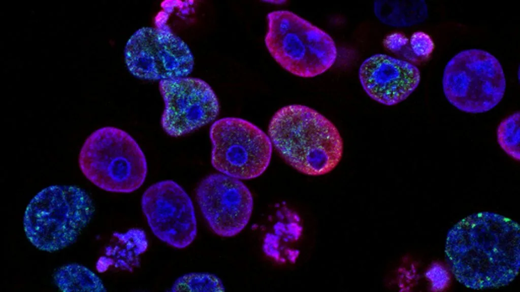
The epigenetic clock represents a mathematical model of biological aging derived from DNA methylation patterns at specific CpG dinucleotides across the human genome. Epigenetic age acceleration strongly correlates with malignant transformation, cancer incidence, and poor prognosis across diverse histopathological tumor subtypes.
- Primary molecular mechanism: Age-associated hypermethylation of specific Polycomb repressive complex 2 (PRC2) target genes and global hypomethylation.
- Diagnostic quantification: Horvath and Hannum multi-tissue clocks utilizing Illumina Infinium Methylation BeadChip arrays.
- Therapeutic intervention: Reversal of aberrant DNA methylation using hypomethylating agents (azacitidine, decitabine) and targeted histone modifiers.
Epigenetic clocks measure biological aging by tracking site-specific DNA methylation status within promoter regions and enhancers throughout the genome. Aberrant acceleration of these methylation clocks distinguishes neoplastic tissues from senescent normal counterparts.
Disruption of homeostatic DNA methyltransferase activity drives widespread genomic instability and transcriptional silencing of crucial tumor suppressor loci. These epigenetic modifications serve as quantitative cellular biomarkers for oncogenic progression and therapeutic stratification.
CLINICAL & LABORATORY DIAGNOSTIC PROTOCOL
- Specimen acquisition: Fresh-frozen or formalin-fixed paraffin-embedded (FFPE) tumor tissue procured with cold ischemia time restricted to less than 30 minutes.
- DNA extraction: Isolation of genomic DNA using silica-column-based kits yielding minimum 500 nanograms of high-purity DNA (A260/A280 ratio between 1.8 and 2.0).
- Bisulfite conversion: Conversion of unmethylated cytosines to uracil using sodium bisulfite chemistry with greater than 99 percent conversion efficiency validated by quantitative control PCR.
Whole-Genome Amplification and Enzymatic Fragmentation
Perform whole-genome amplification on 200 nanograms of bisulfite-converted DNA followed by enzymatic fragmentation using calibrated endonuclease digestion reagents. Incubate reaction mixtures at 37°C for exactly 60 minutes to achieve optimal fragment length distributions between 300 and 600 base pairs.
Hybridization to Illumina Infinium Methylation BeadChip
Denature fragmented DNA samples at 95°C for 20 minutes and apply samples to the Illumina Infinium MethylationEPIC v2.0 BeadChip array. Incubate hybridization chambers in a rotary hybridization oven at 48°C for 16 to 24 hours.
Single-Base Extension and Fluorescence Staining
Execute single-base extension utilizing labeled nucleotides and multi-layered fluorescent staining reagents within an automated Tecan Freedom EVO fluidics workstation. Wash arrays stringently using high-stringency wash buffers at 45°C to eliminate non-specific probe binding.
Array Scanning and Bioinformatic Epigenetic Clock Calculation
Scan processed BeadChips using the Illumina iScan System to capture high-resolution dual-color fluorescence intensities. Extract raw methylation beta values using GenomeStudio software and apply R-based algorithms to compute Horvath, Hannum, and PhenoAge epigenetic age metrics.
1. Pathophysiology, Etiology & Cellular Carcinogenesis
Epigenetic drift during physiological aging introduces stochastic errors in DNA maintenance methylation catalyzed by **DNMT1**, **DNMT3A**, and **DNMT3B**. In oncogenesis, these stochastic alterations converge into distinct methylation signatures that silence critical apoptotic and DNA repair regulators.
Promoter hypermethylation frequently targets CpG islands associated with tumor suppressor genes, whereas global genomic hypomethylation induces chromosomal instability. This dual epigenetic dysregulation creates a permissive cellular environment for clonal evolution and malignant transformation.
The tumor microenvironment actively secretes paracrine factors that accelerate epigenetic aging in adjacent stromal fibroblasts and infiltrating immune cells. This cancer-induced epigenetic remodeling supports tumor immune evasion, angiogenesis, and extracellular matrix degradation.
2. Oncogenic Signal Transduction Pathways & Genomic Alterations
Aberrant epigenetic silencing targets key nodes within the **PI3K-AKT-mTOR** and **RAS-RAF-MEK-ERK** signal transduction pathways. Loss of negative regulators through promoter hypermethylation leads to ligand-independent receptor tyrosine kinase activation and sustained cellular proliferation.
Mutations in epigenetic modifier genes such as **IDH1**, **IDH2**, and **ARID1A** generate oncometabolites that competitively inhibit **TET** family dioxygenases. This enzymatic blockade prevents active DNA demethylation, maintaining cells in an undifferentiated, proliferative state.
Cross-talk between histone deacetylases and DNA methyltransferases solidifies repressive chromatin states at lineage-specific promoters. Bypass signaling cascades emerge when sustained epigenetic silencing circumvents targeted inhibition of upstream kinases.
3. Histopathological Grading, Morphological Subtypes & Differential Diagnosis
Epigenetic clock acceleration correlates with higher histopathological grade, increased mitotic index, and advanced architectural disorganization across carcinomas and sarcomas. Pathological evaluation reveals prominent nuclear pleomorphism and dense heterochromatin redistribution in biologically older tumors.
Diagnostic immunophenotyping utilizes surrogate markers of epigenetic dysregulation, including loss of **ARID1A** or aberrant overexpression of **EZH2**. These immunohistochemical findings complement DNA methylation profiling to refine histopathological classification.
Differential diagnosis requires distinguishing primary hypermethylator phenotypes from secondary metastatic lesions displaying distinct tissue-of-origin methylation signatures. Integration of genome-wide methylation classifiers resolves ambiguities in poorly differentiated neoplasms.
4. Targeted Therapeutics, Landmark Phase III Trials & Resistance Mechanics
Pharmacological targeting of epigenetic aberrations relies on DNA methyltransferase inhibitors such as **azacitidine** and **decitabine**, alongside selective **EZH2** inhibitors like **tazemetostat**. These agents reactivate silenced tumor suppressor genes by inducing genome-wide demethylation and chromatin relaxation.
Landmark clinical trials, including pivotal Phase III evaluations in myelodysplastic syndromes and acute myeloid leukemia, demonstrate improved overall survival and complete remission rates with hypomethylating regimens. Combination strategies incorporating immune checkpoint inhibitors are currently under clinical investigation.
Resistance to epigenetic therapies often arises through upregulation of drug efflux transporters or secondary mutations in metabolic enzymes that process cytidine analogs. Novel bivalent inhibitors targeting both histone readers and DNA methyltransferases are engineered to overcome these resistance hurdles.
5. Molecular Staging, Minimal Residual Disease (ctDNA) & Prognostic Stratification
Integration of epigenetic clock metrics with the **AJCC 8th edition** TNM staging system provides superior prognostic granularity compared to clinicopathological parameters alone. Biological age acceleration consistently predicts diminished overall survival and accelerated disease progression across solid tumors.
Liquid biopsy surveillance employing circulating tumor DNA (**ctDNA**) methylation profiling allows non-invasive detection of minimal residual disease. Quantitative monitoring of specific hypermethylated promoter sequences detects recurrence significantly ahead of radiographic imaging.
Future clinical oncology paradigms utilize personalized epigenetic aging signatures to tailor therapeutic intensity, sparing chemo-naive patients excessive toxicity while escalating intervention in biologically aggressive, rapidly aging tumors.