Epithelial Dysplasia and Pre-Invasive Cellular Pathology: Molecular Signatures, Histological Grading, and Malignant Transformation
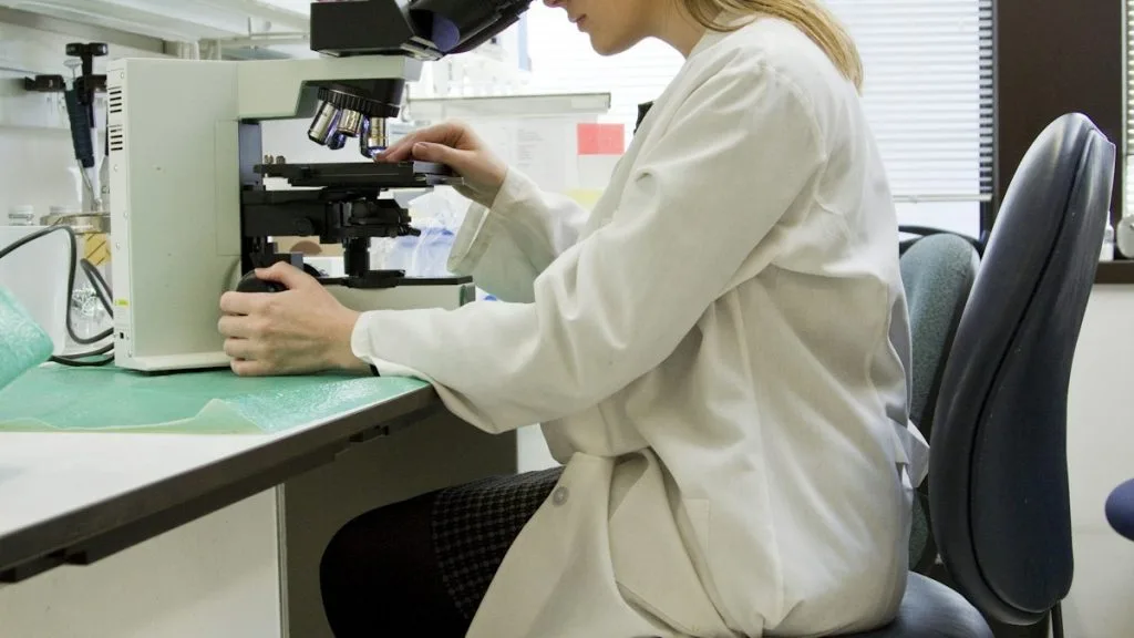
Epithelial dysplasia represents a disordered, pre-malignant cellular proliferation characterized by architectural disorganization and cytological atypia without basement membrane penetration. This cellular pathology state carries a variable probability of transitioning to invasive squamous cell carcinoma or adenocarcinoma depending on anatomical site and genomic instability.
- Primary diagnostic hallmark: Loss of cellular maturation, nuclear pleomorphism, hyperchromasia, and aberrant mitotic figures restricted by the intact basement membrane.
- Molecular driver cascade: Inactivation of tumor suppressor genes **TP53** and **CDKN2A** coupled with telomerase reactivation and chromosomal instability.
- Clinical intervention threshold: High-grade dysplasia demands immediate endoscopic or surgical eradication to prevent invasive carcinoma progression.
Epithelial dysplasia manifests across diverse mucosal and glandular surfaces including the cervix, esophagus, bronchial tree, and gastrointestinal tract. Persistent chronic inflammation, oncogenic viral infections such as high-risk human papillomavirus genotypes 16 and 18, and environmental genotoxins drive this pre-malignant clonal expansion.
The transition from normal epithelium to low-grade and high-grade dysplasia involves progressive accumulation of somatic mutations and epigenetic silencing. Disruption of normal cellular polarity and unchecked proliferation create a distinct morphological phenotype identifiable through standard hematoxylin and eosin staining.
CLINICAL & LABORATORY DIAGNOSTIC PROTOCOL
- Specimen acquisition: Endoscopic mucosal biopsies or excisional surgical specimens fixed immediately in 10% neutral-buffered formalin (NBF) with a cold ischemia time strictly under 60 minutes.
- Tissue processing: Formalin fixation maintained for 24 to 48 hours at room temperature, followed by automated paraffin dehydration and embedding into paraffin blocks.
- Control standards: Internal normal epithelial elements and dedicated external positive control tissue blocks validated for target immunohistochemical stains run in parallel.
Tissue Deparaffinization & Heat-Induced Epitope Retrieval (HIER)
Bake paraffin sections at 60°C for 60 minutes, deparaffinize through three changes of xylene (5 minutes each), and rehydrate via graded ethanols (100%, 95%, 70%) to distilled water. Perform HIER using a high-pH EDTA buffer (pH 9.0) at 97°C for 20 minutes inside an automated pretreatment module.
Endogenous Enzyme Block & Primary Antibody Incubation
Block endogenous peroxidase activity with 3% hydrogen peroxide for 10 minutes at room temperature, followed by rinsing in Tris-buffered saline with 0.05% Tween-20 (TBST). Apply primary monoclonal antibodies targeting **p16** (clone E6H4) or **Ki-67** (clone MIB-1) at pre-titrated optimal dilutions for 30 minutes.
Polymer Detection System & Chromogen Application
Apply a horseradish peroxidase-conjugated secondary goat anti-rabbit/mouse polymer detection reagent for 30 minutes at room temperature. Wash thoroughly in TBST, then apply freshly prepared 3,3-diaminobenzidine (DAB) chromogen substrate solution for exactly 5 minutes.
Counterstaining, Dehydration & Diagnostic Scoring
Counterstain slides with Mayer hematoxylin for 60 seconds, blue in running tap water, dehydrate through ascending ethanols, clear in xylene, and apply permanent synthetic mounting medium. Evaluate architectural disarray, nuclear-to-cytoplasmic ratio, and mitotic distribution under light microscopy.
1. Pathophysiology, Etiology & Cellular Carcinogenesis
Epithelial dysplasia arises from sustained cellular injury and chronic regenerative stimuli that foster genomic instability. Clonal populations acquire survival advantages through telomere attrition evasion and persistent evasion of apoptotic signaling networks.
Epigenetic dysregulation, notably CpG island promoter hypermethylation of tumor suppressor loci such as **CDKN2A**, silences vital cell cycle checkpoints. This molecular reprogramming abolishes contact inhibition and drives architectural crowding.
The surrounding stroma undergoes co-evolution, characterized by myofibroblastic activation, altered extracellular matrix deposition, and pro-angiogenic factor release. This altered microenvironment facilitates pre-malignant persistence and eventual invasion.
2. Oncogenic Signal Transduction Pathways & Genomic Alterations
Genomic sequencing of dysplastic lesions frequently reveals loss-of-function mutations in the **TP53** tumor suppressor gene located on chromosome 17p13.1. This aberration compromises DNA damage response checkpoints and permits propagation of chromosomal abnormalities.
Concurrently, hyperactivation of the **PI3K-AKT-mTOR** pathway promotes anabolic metabolism and macromolecular synthesis required for rapid cellular division. Upregulation of receptor tyrosine kinases further amplifies downstream proliferative signals.
Secondary alterations in the **RAS-RAF-MEK-ERK** cascade drive sustained transcriptional activation of cyclins. These intracellular alterations collectively abolish normal differentiation cues and lock cells into an immature proliferative state.
3. Histopathological Grading, Morphological Subtypes & Differential Diagnosis
Histological evaluation classifies dysplasia into low-grade and high-grade categories based on the extent of architectural atypia. Low-grade dysplasia displays basaloid expansion confined to the lower third of the epithelium, retaining nuclear polarity.
High-grade dysplasia exhibits severe architectural disorganization spanning the middle and upper thirds of the mucosal thickness, accompanied by prominent nucleoli and atypical mitotic figures. The basement membrane remains entirely intact.
Differential diagnosis requires careful exclusion of reactive epithelial atypia secondary to acute inflammation, mucosal erosion, and regenerative hyperplasia. Immunohistochemical stains for **p53** aberrancy and elevated **Ki-67** indices resolve ambiguous cases.
4. Targeted Therapeutics, Landmark Phase III Trials & Resistance Mechanics
Management of localized dysplasia relies on endoscopic eradication therapies, including endoscopic mucosal resection and radiofrequency ablation. These interventions eradicate pre-malignant clones before invasive transformation occurs.
Landmark clinical trials such as the pivotal gastrointestinal endoscopic surveillance and ablation studies demonstrate durable complete eradication rates exceeding 85% for high-grade Barrett esophagus dysplasia. Systemic chemotherapy is reserved for confirmed invasive disease.
Resistance to local ablative therapies often stems from buried dysplastic glands beneath regenerated squamous epithelium. Advanced multi-modality endoscopic mapping and surveillance protocols mitigate recurrence risks.
5. Molecular Staging, Minimal Residual Disease (ctDNA) & Prognostic Stratification
Integration of conventional AJCC 8th edition Tis staging with molecular sequencing profiles enhances risk stratification for high-risk dysplastic lesions. Genomic loss of heterozygosity panels quantify progression probabilities.
Emerging surveillance paradigms utilize circulating tumor DNA and ultra-deep sequencing of mucosal brushings to detect minimal residual genomic aberrations. These assays identify early clonal recurrence prior to overt morphological relapse.
Prognostic stratification models incorporate patient age, lesion length, and multiplex genomic instability scores to guide surveillance intervals. This precise approach optimizes clinical management and prevents progression to advanced metastatic disease.