ESR1 Ligand-Binding Domain Mutations in Endocrine-Resistant Breast Carcinoma: Molecular Pathology, Genomic Evolution & Targeted Therapeutics
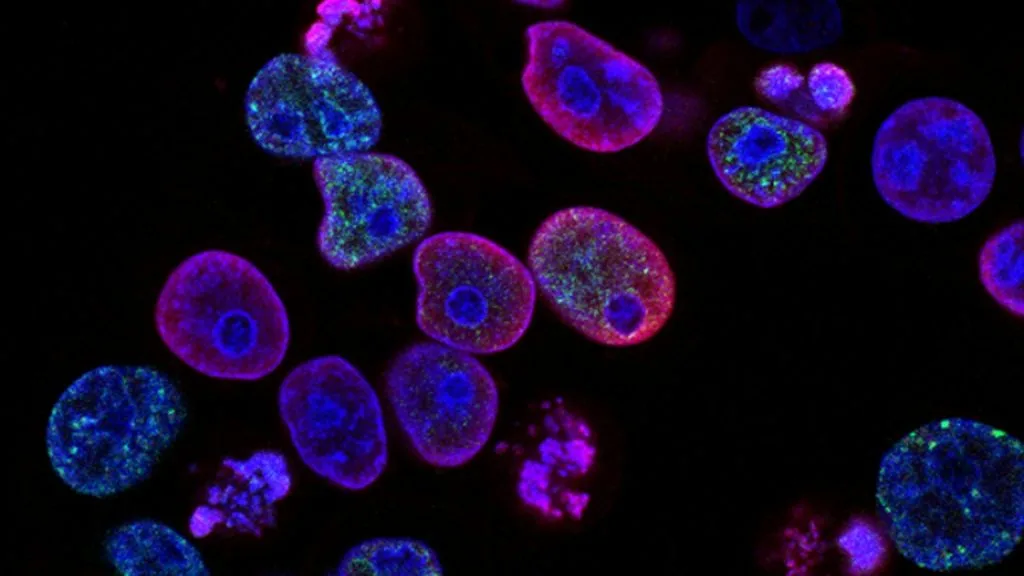
Estrogen receptor 1 (**ESR1**) ligand-binding domain mutations drive acquired endocrine resistance in metastatic estrogen receptor-positive breast carcinoma. These activating missense alterations occur predominantly following prolonged aromatase inhibitor exposure, locking the receptor in a constitutively active transcriptional conformation.
- Primary molecular driver: Activating missense mutations in the **ESR1** ligand-binding domain (codons 530-547).
- First-line standard of care for ESR1-mutated metastatic disease: Oral selective estrogen receptor degrader **elacestrant** or **fulvestrant** combination regimens.
- Primary mechanism of resistance: Ligand-independent constitutive coactivator recruitment and loss of aromatase inhibitor suppression.
Acquired endocrine resistance represents a formidable clinical barrier in the management of hormone receptor-positive metastatic breast carcinoma. Approximately forty percent of patients previously treated with adjuvant or metastatic aromatase inhibitor therapy develop secondary **ESR1** mutations.
These structural alterations convert the estrogen receptor alpha protein into an agonist conformation without requiring ligand binding. Consequently, downstream transcriptional activation persists unabated despite profound estrogen deprivation induced by letrozole, anastrozole, or exemestane.
CLINICAL & LABORATORY DIAGNOSTIC PROTOCOL
- Specimen acquisition: Archival formalin-fixed paraffin-embedded tissue block or cell-free circulating tumor DNA (ctDNA) liquid biopsy extracted from 10 mL peripheral blood collected in Streck Cell-Free DNA BCT tubes.
- Tissue processing: Formalin fixation time strictly calibrated to 24-48 hours in 10% neutral-buffered formalin at room temperature; microtome sectioning at 5-micron thickness for molecular assays.
- Control standards: Wild-type and mutant control cell line DNA spiked into analytical runs to validate limit of detection for droplet digital PCR or next-generation sequencing assays.
Genomic DNA Extraction & Quantification
Isolate genomic DNA from microdissected tumor areas containing at least 20% neoplastic cellularity using automated silica-membrane spin-column extraction kits. Quantify double-stranded DNA yield via fluorometric assays to ensure input requirements for targeted sequencing panels are met.
Targeted Amplicon Library Preparation
Perform multiplex polymerase chain reaction amplification targeting **ESR1** exons 8 and 9, encompassing the ligand-binding domain codons 530 through 547. Incorporate standard Illumina or Ion Torrent adapter sequences and unique molecular identifiers during library construction.
Next-Generation Sequencing (NGS) or Digital PCR
Sequence prepared libraries on a validated high-throughput sequencing platform achieving a minimum mean depth of coverage of 1000x across targeted **ESR1** loci. Alternatively, utilize droplet digital PCR assays specifically tailored for **Y537S** and **D538G** hot spot detection.
Bioinformatic Pipeline & Variant Calling
Align raw sequencing reads to the GRCh38 human reference genome using Burrows-Wheeler Aligner software. Filter somatic variants using somatic mutation callers, applying a strict variant allele frequency threshold of greater than 2 percent for tissue and 0.5 percent for ctDNA.
1. Pathophysiology, Etiology & Cellular Carcinogenesis
Endocrine resistance emerges through Darwinian selection of minor pre-existing subclones or de novo mutagenesis during aromatase inhibitor therapy. Estrogen deprivation creates intense selective pressure favoring **ESR1** missense mutations within the ligand-binding domain.
Epigenetic dysregulation and histone modification cooperate with genomic alterations to sustain uninhibited cellular proliferation. Cyclin D-CDK4/6 complexes remain constitutively phosphorylated, driving escape from cell cycle arrest pathways.
The tumor microenvironment recruits immunosuppressive myeloid-derived suppressor cells and cancer-associated fibroblasts. This stroma secretes paracrine growth factors that activate bypass receptor tyrosine kinase pathways.
2. Oncogenic Signal Transduction Pathways & Genomic Alterations
Wild-type estrogen receptor alpha requires agonist binding to induce a structural shift in helix 12, permitting coactivator recruitment. **ESR1** mutations such as **Y537S** and **D538G** stabilize helix 12 in an active agonist position without ligand binding.
Constitutive nuclear receptor activation stimulates transcription of downstream genes regulating cell cycle progression and survival. Crosstalk with the **PI3K-AKT-mTOR** and **MAPK** pathways amplifies proliferative signaling and therapeutic resistance.
Secondary concurrent genomic alterations, including **PIK3CA** helical or kinase domain mutations and **CCND1** amplification, frequently coexist with **ESR1** mutations. These co-alterations reinforce evasion of endocrine blockade and necessitate combinatorial drug inhibition.
3. Histopathological Grading, Morphological Subtypes & Differential Diagnosis
Endocrine-resistant metastatic breast carcinomas predominantly exhibit invasive ductal carcinoma of no special type morphology. High nuclear grade, prominent nucleoli, and brisk mitotic activity characterize these chemo- and endocrine-resistant lesions.
Diagnostic immunohistochemistry demonstrates retained nuclear expression of **ER**, variable **PR** attenuation or loss, and negative **HER2** overexpression. Concomitant loss of **GATA3** or aberrant cytokeratin profiles may accompany advanced phenotypic dedifferentiation.
Differential diagnosis requires distinguishing acquired endocrine resistance from second primary malignancies or poorly differentiated metastatic mimics. Comprehensive immunohistochemical panels including **GATA3**, **Mammaglobin**, and **GCDFP-15** confirm mammary primary origin.
4. Targeted Therapeutics, Landmark Phase III Trials & Resistance Mechanics
First-generation selective estrogen receptor modulators like tamoxifen fail against **ESR1** mutants due to partial agonist activity. Selective estrogen receptor degraders such as **fulvestrant** induce receptor degradation but possess pharmacokinetic limitations.
The landmark Phase III EMERALD clinical trial evaluated oral **elacestrant** versus standard endocrine monotherapy in previously treated metastatic breast cancer. **Elacestrant** demonstrated significantly improved progression-free survival in patients harboring confirmed **ESR1** mutations.
Next-generation covalent oral SERDs and complete estrogen receptor antagonists are currently investigated in clinical trials to overcome emerging tertiary resistance mutations. Combining novel degraders with **CDK4/6 inhibitors** or **mTOR inhibitors** represents an active frontier.
5. Molecular Staging, Minimal Residual Disease (ctDNA) & Prognostic Stratification
Integration of **AJCC 8th edition** TNM staging parameters with circulating genomic biomarkers refines prognostic models. Detection of mutant **ESR1** allelic fractions in serial liquid biopsies correlates with impending clinical progression.
Circulating tumor DNA surveillance provides dynamic monitoring of minimal residual disease and early treatment failure detection. Quantitative tracking of mutant allele fractions informs timely therapeutic switching before radiographic manifestation.
Patients with detectable **ESR1** mutations experience significantly shorter progression-free survival on subsequent endocrine therapies. Incorporating molecular profiling into routine oncology workflows guides precision treatment algorithms for advanced breast cancer.