Genomic Instability in Neoplastic Progression: DNA Damage Repair Deficiencies, Chromosomal Instability & Synthetic Lethality
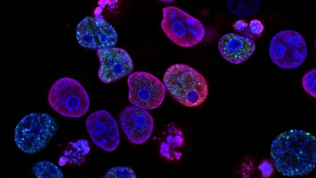
Genomic instability is a hallmark of malignancy characterized by a high rate of alterations in the genome, spanning nucleotide mutations to gross chromosomal aberrations. It drives intratumoral heterogeneity, therapeutic resistance, and aggressive clinical phenotypes across solid and hematologic tumors.
- Primary driver mechanism: Impairment of DNA damage response (DDR) networks including homologous recombination and mismatch repair.
- Therapeutic exploitation: Clinical utilization of PARP inhibitors to induce synthetic lethality in HR-deficient malignancies.
- Immunotherapeutic vulnerability: Hypermutated phenotypes resulting from DNA repair failure generate neoantigens responsive to immune checkpoint inhibition.
Genomic instability manifests across diverse human malignancies, correlating with advanced stage, high histological grade, and dismal clinical outcomes. It arises from persistent replication stress, telomere attrition, and defective mitotic checkpoints.
The cellular consequence of this instability is accelerated mutagenesis, empowering subclonal populations to adapt under selective therapeutic pressures. Elucidating these repair defects provides actionable targets for molecularly tailored interventions.
CLINICAL & LABORATORY DIAGNOSTIC PROTOCOL
- Specimen acquisition: Fresh surgical resection or core needle biopsy fixed in 10% neutral-buffered formalin (NBF) within 15 minutes of devascularization to preserve DNA integrity.
- Tissue processing: Controlled formalin fixation for 24 to 48 hours at room temperature; paraffin embedding followed by microtome sectioning at 4-micron thickness onto superfrost plus slides.
- Control standards: Wild-type and known mutant tissue controls processed alongside clinical specimens to validate analytical sensitivity and specificity.
DNA Extraction & Quality Assessment
Isolate genomic DNA from macrodissected formalin-fixed paraffin-embedded (FFPE) tumor tissue using silica-column extraction kits. Quantify double-stranded DNA yield via fluorometry and assess fragmentation status using capillary electrophoresis.
Next-Generation Sequencing (NGS) Library Preparation
Perform enzymatic DNA fragmentation, end-repair, A-tailing, and adapter ligation using target enrichment panels covering core DNA repair genes (**BRCA1**, **BRCA2**, **ATM**, **PALB2**). Amplify libraries via polymerase chain reaction using unique molecular identifiers (UMIs) to suppress PCR artifacts.
Bioinformatic Pipeline Execution & Variant Calling
Align FASTQ reads to the GRCh37/hg19 reference genome using BWA-MEM, mark duplicates, and recalibrate base quality scores. Execute somatic variant calling algorithms (Mutect2, Strelka2) to identify single nucleotide variants, small insertions/deletions, and copy number alterations.
Genomic Scar Scoring & Clinical Reporting
Calculate genome-wide loss of heterozygosity (LOH), telomeric allelic imbalance (TAI), and large-scale state transitions (LST) to compute an aggregate **HRD score**. Format final molecular pathology report detailing pathogenic mutations, variant allele frequencies, and companion diagnostic classifications.
1. Pathophysiology, Etiology & Cellular Carcinogenesis
Genomic instability originates from the breakdown of surveillance mechanisms that normally maintain structural and numerical chromosomal integrity. Mutations in **TP53** abrogate the G1/S cell cycle checkpoint, permitting cells with damaged DNA to enter S phase and accumulate mutations.
Epigenetic silencing through promoter hypermethylation of DNA repair genes, such as **MLH1** or **MGMT**, further compromises genomic fidelity. This progressive mutational load fuels Darwinian selection within the tumor microenvironment, yielding aggressive cellular clones.
Chronic replication stress induces stalled replication forks, which if unresolved, collapse into double-strand breaks. The resulting chromosomal aberrations drive structural rearrangements, including translocations, deletions, and oncogene amplifications.
2. Oncogenic Signal Transduction Pathways & Genomic Alterations
Defects in homologous recombination repair (**HRR**) force cancer cells to rely on error-prone non-homologous end joining (**NHEJ**). This error-prone pathway introduces microhomologies and random insertions, exacerbating karyotypic complexity.
Concurrently, hyperactivation of the **PI3K-AKT-mTOR** pathway alters metabolic cellular states to sustain high rates of DNA replication. Upregulated checkpoint kinases **ATR** and **CHK1** are persistently engaged to manage elevated baseline replication stress.
Bypass signaling cascades compensate for stalled DNA repair machineries by activating alternative survival pathways such as **RAS-RAF-MEK-ERK**. This molecular rewiring sustains unchecked cellular proliferation despite lethal genomic damage.
3. Histopathological Grading, Morphological Subtypes & Differential Diagnosis
Tumors characterized by high chromosomal instability exhibit marked nuclear pleomorphism, atypical mitotic figures, and bizarre multinucleated giant cells. Histological grading reflects this architectural anarchy, often classifying affected neoplasms as high-grade or poorly differentiated.
Immunohistochemical panels evaluating loss of mismatch repair proteins (**MLH1**, **MSH2**, **MSH6**, **PMS2**) serve as initial screening surrogates for microsatellite instability. Retained nuclear staining acts as an internal positive control, whereas absent expression indicates underlying mismatch repair deficiency.
The differential diagnosis requires distinguishing primary poorly differentiated carcinomas from metastatic deposits exhibiting high-grade features. Comprehensive immunohistochemical profiling helps delineate lineage-specific markers from secondary stromal contaminants.
4. Targeted Therapeutics, Landmark Phase III Trials & Resistance Mechanics
The clinical exploitation of genomic instability relies on the concept of synthetic lethality induced by poly (ADP-ribose) polymerase (**PARP**) inhibitors like **olaparib** and **niraparib**. Inhibiting base excision repair in tumors with pre-existing **BRCA1/2** mutations causes catastrophic double-strand break accumulation and apoptotic cell death.
Landmark Phase III clinical trials, including **SOLO1** and **PAOLA-1**, established the profound progression-free survival benefit of maintenance **olaparib** in advanced ovarian carcinoma harboring homologous recombination deficiency. Similarly, the **OlympiA** trial demonstrated invasive disease-free survival improvements in adjuvant **olaparib** administration for high-risk **BRCA**-mutant breast cancer.
Acquired resistance to **PARP** inhibitors frequently emerges via secondary reversion mutations in **BRCA1** or **BRCA2** that restore the reading frame and homologous recombination function. Combination strategies incorporating immune checkpoint inhibitors such as **pembrolizumab** aim to overcome this resistance by leveraging neoantigen loads.
5. Molecular Staging, Minimal Residual Disease (ctDNA) & Prognostic Stratification
Integration of genomic instability metrics into the AJCC 8th edition staging framework refines prognostic stratification beyond traditional clinicopathologic parameters. Tumors harboring high genomic scar scores demonstrate distinct clinical trajectories and heightened sensitivity to DNA-damaging cytotoxic agents.
Liquid biopsy platforms employing circulating tumor DNA (**ctDNA**) tracking detect minimal residual disease and monitor subclonal evolution in real time. Serial quantification of mutant allele fractions provides early detection of recurrence prior to radiologic manifestation.
Prospective clinical paradigms increasingly utilize molecular signatures of genomic instability to guide personalized treatment de-escalation or escalation protocols. This precision oncology approach maximizes therapeutic efficacy while minimizing systemic toxicities in molecularly selected patient cohorts.