HER2/neu (ERBB2) Amplification & Trastuzumab Deruxtecan Antibody-Drug Conjugate
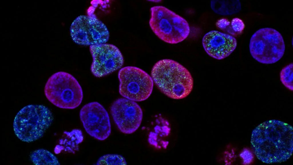
HER2/neu amplification drives aggressive tumorigenesis via ERBB2 gene overexpression, warranting precise diagnostic evaluation and antibody-drug conjugate therapy. Trastuzumab deruxtecan delivers a potent topoisomerase I inhibitor payload directly to HER2-expressing tumor cells.
- Primary molecular alteration: ERBB2 gene amplification and protein overexpression driving oncogenic signaling.
- First-line therapeutic standard: Anti-HER2 monoclonal antibodies combined with cytotoxic chemotherapy.
- Antibody-drug conjugate efficacy: Trastuzumab deruxtecan demonstrates profound survival benefit in HER2-positive and HER2-low metastatic settings.
HER2/neu overexpression occurs in approximately 15 to 20 percent of invasive breast carcinomas and a distinct subset of gastroesophageal, pulmonary, and gynecological malignancies. Patients presenting with this oncogenic driver frequently exhibit high histologic grade, accelerated cellular proliferation rates, and heightened propensity for visceral metastasis.
The **ERBB2** proto-oncogene encodes a 185-kilodalton transmembrane receptor tyrosine kinase belonging to the epidermal growth factor receptor family. Gene amplification leads to dense receptor clustering on the plasma membrane, triggering ligand-independent activation of downstream mitogenic pathways.
CLINICAL & LABORATORY DIAGNOSTIC PROTOCOL
- Specimen acquisition: Core needle biopsy or surgical resection fixed in 10 percent neutral-buffered formalin (NBF) within 30 minutes of devascularization (cold ischemia time strictly under 1 hour).
- Tissue processing: Formalin fixation duration strictly calibrated to 6 to 72 hours at room temperature; paraffin embedding followed by 4-micron microtome sectioning on positively charged adhesive slides.
- Control standards: On-slide external positive control breast carcinoma tissue with known IHC 3+ amplification and validated negative tissue controls run in parallel with every analytical batch.
Tissue Deparaffinization & Heat-Induced Epitope Retrieval (HIER)
Bake slides at 60°C for 60 minutes, deparaffinize through sequential xylene baths (3 changes of 5 minutes each), and rehydrate through graded ethanols (100 percent, 95 percent, 70 percent) to deionized water. Perform HIER using a high-pH tris-EDTA buffer (pH 9.0) at 97°C for 20 minutes in an automated slide processing module.
Endogenous Peroxidase Block & Primary Antibody Incubation
Quench endogenous peroxidase activity by incubating sections in 3 percent hydrogen peroxide for 10 minutes at room temperature. Apply optimized anti-HER2 primary monoclonal antibody (clone 4B5 or CB11) at validated clinical dilution for 32 minutes in a humidified chamber.
Polymer Detection System & DAB Chromogen Visualization
Rinse sections thoroughly in Tris-buffered saline with 0.05 percent Tween-20 (TBST). Apply horseradish peroxidase (HRP)-conjugated anti-rabbit/mouse secondary polymer reagent for 16 minutes, followed by incubation with freshly prepared 3,3-diaminobenzidine (DAB) substrate-chromogen solution for 8 minutes to generate a brown precipitate.
Counterstaining, Dehydration & Quality Control Scoring
Counterstain nuclei with Mayer hematoxylin for 4 minutes, blue in running tap water, dehydrate through graded ethanols, clear in xylene, and coverslip using permanent non-aqueous mounting medium. Evaluate complete, intense circumferential membrane staining in greater than 10 percent of invasive tumor cells for an IHC 3+ score.
1. Pathophysiology, Etiology & Cellular Carcinogenesis
Cell transformation driven by **ERBB2** amplification originates from chromosomal instability and localized DNA replication errors on chromosome 17q12-q21. This genomic aberration results in hundreds of copies of the **ERBB2** locus, shifting cellular homeostasis toward unchecked proliferation.
Epigenetic dysregulation and promoter modifications further enhance transcriptional output, driving excessive receptor density on the plasma membrane. Evasion of cell cycle checkpoints allows continuously dividing malignant clones to bypass normal apoptotic pathways.
The surrounding tumor microenvironment adapts to this aggressive signaling via dense desmoplastic stroma and pro-angiogenic cytokine release. Hypoxic zones within the tumor core stimulate vascular endothelial growth factor production, facilitating local invasion and hematogenous dissemination.
2. Oncogenic Signal Transduction Pathways & Genomic Alterations
Upstream receptor activation occurs through ligand-independent homodimerization or heterodimerization with other members of the ERBB family, particularly **HER3** (**ERBB3**). This physical interaction induces conformational shifts that stimulate intrinsic tyrosine kinase domain transautophosphorylation.
Phosphorylated intracellular tyrosine residues dock specific adaptor proteins, initiating robust downstream signaling cascades through the **RAS-RAF-MEK-ERK** and **PI3K-AKT-mTOR** pathways. Persistent activation of these cascades drives accelerated transcription of genes governing cell division, survival, and motility.
Secondary alterations or feedback loops can induce resistance to targeted blockade by activating alternate receptor tyrosine kinases such as **EGFR** or **MET**. These bypass signaling networks maintain downstream survival signals despite effective primary receptor inhibition.
3. Histopathological Grading, Morphological Subtypes & Differential Diagnosis
Architectural growth patterns in HER2-positive carcinomas frequently display solid, cribriform, or micropapillary features with marked nuclear atypia and prominent nucleoli. High mitotic indices and frequent atypical mitotic figures correspond to Nottingham histologic grade 3 morphology.
Diagnostic immunohistochemical panels incorporate **ER**, **PR**, and **HER2**, alongside lineage-specific markers to establish precise tumor classification and therapeutic eligibility. In ambiguous cases, dual-probe fluorescence in situ hybridization (FISH) confirms gene amplification by measuring the **ERBB2** to **CEP17** ratio.
Differential diagnosis requires distinguishing primary HER2-amplified adenocarcinomas from metastatic lesions, lobular carcinomas, and benign atypical ductal hyperplasias. Precise morphological assessment prevents misclassification and ensures appropriate deployment of targeted antibody-drug conjugates.
4. Targeted Therapeutics, Landmark Phase III Trials & Resistance Mechanics
Therapeutic strategies rely on monoclonal antibodies, tyrosine kinase inhibitors, and antibody-drug conjugates designed to exploit **HER2** dependency. **Trastuzumab deruxtecan** (**T-DXd**) delivers a potent membrane-permeable topoisomerase I inhibitor payload intracellularly via a stable cleavable tetrapeptide-based linker.
Landmark clinical trials such as DESTINY-Breast03 and DESTINY-Breast04 established **trastuzumab deruxtecan** as a transformative standard of care, demonstrating unprecedented progression-free survival advantages over standard chemotherapy in both high-expressing and HER2-low cohorts. Overall survival curves show marked separation favoring the antibody-drug conjugate.
Resistance mechanisms to antibody-drug conjugates include down-regulation of surface **HER2** expression, impaired internalization kinetics, and multidrug resistance transporter upregulation. Ongoing clinical evaluations investigate rational combination regimens with immune checkpoint inhibitors to overcome refractory disease phenotypes.
5. Molecular Staging, Minimal Residual Disease (ctDNA) & Prognostic Stratification
Integration of AJCC 8th edition anatomic TNM parameters with **HER2** biomarker status provides a comprehensive prognostic framework for clinical decision-making. Pathologic complete response following neoadjuvant systemic therapy correlates strongly with superior long-term disease-free survival.
Liquid biopsy surveillance utilizing circulating tumor DNA (**ctDNA**) allows non-invasive tracking of minimal residual disease and emergence of resistance mutations. Detecting clonal evolution prior to radiologic recurrence enables timely adjustment of systemic regimens.
Prognostic stratification models incorporate genomic instability scores and multi-gene expression panels to tailor surveillance intervals and optimize adjuvant treatment duration. These precision oncology paradigms continue to refine survival outcomes across diverse clinical populations.