Homeobox Protein NANOG in Cellular Pathology: Embryonic Pluripotency Networks, Cancer Stem Cell Maintenance & Therapeutic Resistance
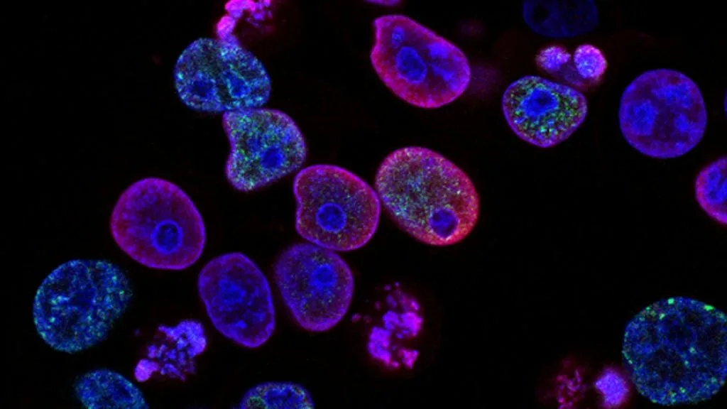
Homeobox protein NANOG is a master transcription factor governing embryonic stem cell pluripotency that undergoes aberrant reactivation in human malignancies. Its dysregulation drives cancer stem cell self-renewal, therapeutic resistance, and aggressive metastatic dissemination.
- Primary molecular function: Transcriptional activation of self-renewal genes alongside POU5F1 and SOX2.
- Oncogenic driver impact: Upregulated expression promotes epithelial-mesenchymal transition and chemotherapy evasion.
- Diagnostic utility: Nuclear immunohistochemical overexpression correlates with advanced tumor stage and poor prognosis.
Aberrant reactivation of embryonic transcriptional networks is a hallmark of aggressive tumorigenesis, cellular dedifferentiation, and poor clinical prognosis. The **NANOG** gene locus on human chromosome 12p13.31 encodes a core transcription factor that maintains pluripotency in embryonic stem cells.
In pathological contexts, **NANOG** expression is hijacked by neoplastic clones to sustain a cancer stem cell phenotype characterized by unlimited self-renewal, evasion of apoptosis, and resistance to standard genotoxic therapies. Unlocking the molecular mechanisms of **NANOG** signaling provides critical diagnostic and therapeutic targets across diverse human cancers.
CLINICAL & LABORATORY DIAGNOSTIC PROTOCOL
- Specimen acquisition: Core needle biopsy or surgical resection fixed in 10% neutral-buffered formalin (NBF) within 30 minutes of devascularization (cold ischemia time < 1 hr).
- Tissue processing: Formalin fixation time strictly calibrated to 24–48 hours at room temperature; paraffin embedding with 4-micron microtome sectioning on positively charged slides.
- Control standards: On-slide external positive control tissue (embryonic testis or seminoma) and validated negative isotype control run in parallel with every analytical batch.
Tissue Deparaffinization & Heat-Induced Epitope Retrieval (HIER)
Bake slides at 60°C for 60 minutes, deparaffinize through sequential xylene baths (3 x 5 min), and rehydrate through graded ethanols (100%, 95%, 70%) to deionized water. Perform HIER in target retrieval solution (pH 9.0 EDTA buffer) at 97°C for 20 minutes in a calibrated PT Link module.
Endogenous Peroxidase Block & Primary Antibody Incubation
Quench endogenous peroxidase activity using 3% hydrogen peroxide for 10 minutes. Apply optimized anti-NANOG primary monoclonal antibody (e.g., clone D73G4) at a calibrated clinical dilution of 1:200 and incubate for 60 minutes at room temperature in a humidified chamber.
Polymer Detection System & DAB Chromogen Visualization
Rinse in Tris-buffered saline with 0.05% Tween-20 (TBST). Apply horseradish peroxidase (HRP)-conjugated anti-rabbit polymer secondary reagent for 30 minutes. Incubate with freshly prepared 3,3-diaminobenzidine (DAB) substrate-chromogen solution for 5-10 minutes until brown precipitates develop.
Counterstaining, Dehydration & Quality Control Scoring
Counterstain nuclei with Mayer hematoxylin for 1–2 minutes, blue in running tap water, dehydrate through graded ethanols, clear in xylene, and coverslip with permanent non-aqueous mounting medium. Score tumor cell nuclear staining under light microscopy using validated scoring algorithm.
1. Pathophysiology, Etiology & Cellular Carcinogenesis
Cellular transformation frequently initiates via the dedifferentiation of somatic epithelial cells into a plastic, embryonic-like state. **NANOG** acts in concert with **OCT4** and **SOX2** to silence lineage-specific differentiation programs and maintain unchecked proliferative capacity.
Epigenetic dysregulation, including promoter hypomethylation of pluripotency loci and histone modifications executed by SWI/SNF complexes, sustains high transcriptional output of **NANOG**. This transcriptional reprogramming unlocks self-renewal pathways previously restricted to the inner cell mass.
Within the tumor microenvironment, hypoxia-inducible factors (**HIF-1alpha**) directly bind to hypoxia-response elements in the **NANOG** promoter. This hypoxic induction enriches the cancer stem cell fraction and drives immunosuppressive stroma formation.
2. Oncogenic Signal Transduction Pathways & Genomic Alterations
Upstream activation of the **JAK2-STAT3** signaling cascade provides constitutive phosphorylation of downstream transcription factors that translocate to the nucleus and activate **NANOG** transcription. Concurrently, **PI3K-AKT-mTOR** pathway hyperactivation stabilizes **NANOG** protein through inhibition of glycogen synthase kinase 3 beta (**GSK3beta**).
Downstream targets of **NANOG** include cell cycle regulators such as cyclin D1 and anti-apoptotic proteins like **BCL-2**, conferring resistance to anoikis and programmed cell death. This network promotes sustained cellular proliferation independent of exogenous growth factor stimulation.
Secondary amplification events at the chromosome 12p13 locus drive supraphysiological levels of **NANOG**, circumventing normal cellular senescence checkpoints and bypassing oncogene-induced senescence.
3. Histopathological Grading, Morphological Subtypes & Differential Diagnosis
Malignancies exhibiting high **NANOG** immunohistochemical expression typically display high-grade morphological features, including prominent nucleoli, high nuclear-to-cytoplasmic ratios, and brisk mitotic activity. Architectural growth patterns often demonstrate solid, poorly differentiated nests with loss of organized glandular or squamous architecture.
Diagnostic evaluation requires a comprehensive immunohistochemical panel incorporating nuclear **NANOG**, **OCT4**, and **SOX2** to confirm the cancer stem cell phenotype, alongside lineage-specific markers to determine the primary site of origin. Pathologists must differentiate primary poorly differentiated carcinomas from metastatic germ cell tumors.
Differential diagnosis includes embryonal carcinoma, high-grade neuroendocrine carcinoma, and melanoma. Nuclear co-localization of **NANOG** and **OCT4** strongly supports embryonal lineage or pluripotent stem cell derivation.
4. Targeted Therapeutics, Landmark Phase III Trials & Resistance Mechanics
Direct pharmacologic inhibition of transcription factors remains challenging, prompting the deployment of upstream signal inhibitors targeting pathways that maintain **NANOG** expression. Small-molecule inhibitors of **JAK2** (e.g., ruxolitinib) and **STAT3** successfully disrupt the transcriptional activation loops driving **NANOG** transcription in refractory solid tumors.
Landmark clinical trials such as the **JAKPOT** and various phase III basket studies evaluating targeted kinase inhibitors demonstrate that suppressing cancer stem cell regulators reduces recurrence rates and sensitizes tumors to cytotoxic chemotherapy. Progression-free survival metrics significantly improve when pluripotency pathways are blocked alongside standard regimens.
Resistance to conventional chemotherapy often emerges through **NANOG**-mediated upregulation of ATP-binding cassette (**ABC**) drug efflux transporters like **ABCG2**. Novel antibody-drug conjugates and bispecific T-cell engagers directed against surface markers co-expressed with **NANOG** are currently under active clinical evaluation.
5. Molecular Staging, Minimal Residual Disease (ctDNA) & Prognostic Stratification
Integration of **NANOG** expression status with AJCC 8th edition TNM staging provides superior prognostic stratification compared to histological grading alone. High nuclear **NANOG** score correlates directly with advanced nodal metastasis and decreased overall survival across multiple solid tumor lineages.
Liquid biopsy surveillance utilizing circulating tumor DNA (**ctDNA**) allows non-invasive tracking of clones harboring pluripotency amplifications. Detection of persisting **NANOG** transcripts in plasma post-resection serves as an early indicator of minimal residual disease and impending relapse.
Prospective clinical paradigms increasingly incorporate stem cell transcription factor profiling into molecular tumor boards to guide personalized therapy selection. Targeting the **NANOG** signaling network represents a critical frontier in overcoming therapeutic resistance and metastatic progression.