Hypervascularity in Solid Neoplasms: Angiogenic Mechanisms, Histopathological Quantification & Anti-Angiogenic Therapies
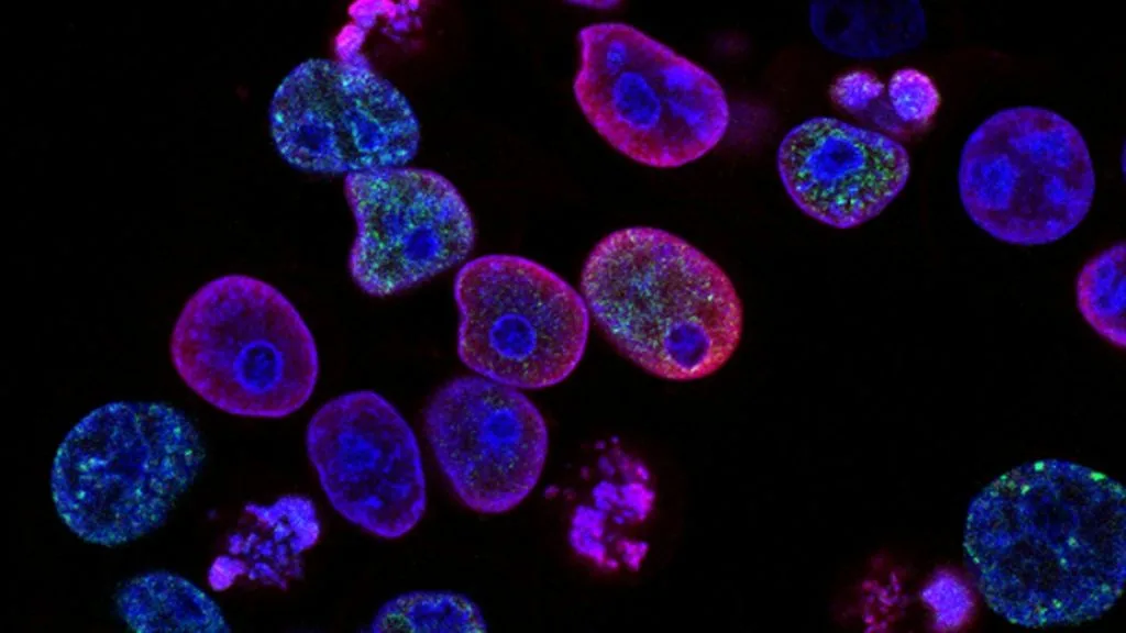
Hypervascularity denotes abnormal, dense microvessel proliferation driven by angiogenic factors within solid tumors. This microstructural alteration dictates aggressive metastatic potential and informs targeted anti-angiogenic therapeutic strategies across diverse oncology indications.
- Primary angiogenic driver: Hypoxia-inducible factor 1-alpha (**HIF1A**) upregulation transcriptionally activating **VEGFA**.
- Pathological quantification: Assessment of intratumoral microvessel density (**MVD**) via endothelial markers **CD31** and **CD34**.
- Therapeutic standard: Monoclonal antibodies neutralizing vascular endothelial growth factor, such as **bevacizumab**, combined with chemotherapy.
Hypervascularity represents a hallmark of aggressive solid malignancies, characterized by tortuous, leaky microvasculature and high endothelial proliferation rates. This phenomenon directly correlates with advanced tumor stage, elevated metastatic recurrence, and poor clinical survival metrics across multiple histotypes.
The pathological basis of tumor hypervascularity stems from an imbalance between pro-angiogenic factors and endogenous inhibitors within the tumor microenvironment. Sustained autocrine and paracrine signaling pathways promote endothelial cell migration, vascular sprouting, and structural remodeling.
CLINICAL & LABORATORY DIAGNOSTIC PROTOCOL
- Specimen acquisition: Core needle biopsy or surgical resection fixed in 10% neutral-buffered formalin (NBF) within 30 minutes of devascularization (cold ischemia time < 1 hr).
- Tissue processing: Formalin fixation time strictly calibrated to 24–48 hours at room temperature; paraffin embedding with 4-micron microtome sectioning on positively charged slides.
- Control standards: On-slide external positive control tissue (tonsil or hemangioma) and validated negative isotype control run in parallel with every analytical batch.
Tissue Deparaffinization & Heat-Induced Epitope Retrieval (HIER)
Bake slides at 60°C for 60 minutes, deparaffinize through sequential xylene baths (3 x 5 min), and rehydrate through graded ethanols (100%, 95%, 70%) to deionized water. Perform HIER in target retrieval solution (pH 9.0 EDTA buffer) at 97°C for 20 minutes in a calibrated PT Link module.
Endogenous Peroxidase Block & Primary Antibody Incubation
Quench endogenous peroxidase activity using 3% hydrogen peroxide for 10 minutes. Apply optimized primary monoclonal antibody targeting **CD31** (clone JC/70A) or **CD34** (clone QBEnd/10) at calibrated clinical dilution and incubate for 30–60 minutes at room temperature in a humidified chamber.
Polymer Detection System & DAB Chromogen Visualization
Rinse in Tris-buffered saline with 0.05% Tween-20 (TBST). Apply horseradish peroxidase (HRP)-conjugated anti-mouse polymer secondary reagent for 30 minutes. Incubate with freshly prepared 3,3-diaminobenzidine (DAB) substrate-chromogen solution for 5-10 minutes until brown precipitates develop.
Counterstaining, Dehydration & Quality Control Scoring
Counterstain nuclei with Mayer hematoxylin for 1–2 minutes, blue in running tap water or 0.08% ammonia water, dehydrate through graded ethanols, clear in xylene, and coverslip with permanent non-aqueous mounting medium. Score microvessel density in hotspots using light microscopy at 200x magnification.
1. Pathophysiology, Etiology & Cellular Carcinogenesis
Intratumor hypoxia acts as the primary biochemical trigger for hypervascularity via stabilization of **HIF1A** and **HIF2A** subunits. Under hypoxic stress, these transcription factors evade proteasomal degradation and translocate to the nucleus to bind hypoxia-response elements.
Epigenetic silencing of endogenous anti-angiogenic genes, such as **THBS1** (thrombospondin-1) and **COL18A1** (endostatin), further tilts the cellular equilibrium toward unchecked neovascularization. Promoter hypermethylation profiles frequently characterize hypervascular tumor subsets.
The resulting tumor microenvironment features erratic blood flow, elevated interstitial fluid pressure, and severe local acidosis. This hostile metabolic niche paradoxically selects for highly invasive cancer cell clones capable of intravasation.
2. Oncogenic Signal Transduction Pathways & Genomic Alterations
Ligand binding of **VEGFA** to endothelial **VEGFR2** triggers receptor dimerization and intrinsic tyrosine kinase autophosphorylation. This initiates downstream signaling cascades including the **PI3K-AKT-mTOR** and **PLCg-PKC** pathways.
Concurrently, the **RAS-RAF-MEK-ERK** pathway is heavily engaged to drive endothelial cell proliferation, survival, and cytoskeletal rearrangement. Cross-talk with fibroblast growth factor (**FGF**) signaling amplifies angiogenic sprouting intensity.
In renal cell carcinoma and specific vascular sarcomas, inactivating mutations of the **VHL** tumor suppressor gene induce constitutive **HIF** activation independently of tissue oxygen tension. This constitutive signaling drives profound baseline hypervascularity.
3. Histopathological Grading, Morphological Subtypes & Differential Diagnosis
Histologically, hypervascular tumors exhibit a dense, disorganized capillary network surrounding malignant nests and cords. Endothelial cells often appear plump, hypertrophic, and mitotically active compared to resting endothelium.
Diagnostic immunohistochemical panels utilizing **CD31**, **CD34**, and **ERG** facilitate accurate microvessel density quantification and endothelial lineage confirmation. Diffuse nuclear and cytoplasmic staining confirms microvascular architectural arrangements.
Differential diagnosis requires distinguishing genuine tumor-induced neovascularization from benign vascular proliferations like hemangiomas, pyogenic granulomas, or vascular malformations. Careful evaluation of atypical cytological features and invasive borders resolves diagnostic ambiguity.
4. Targeted Therapeutics, Landmark Phase III Trials & Resistance Mechanics
Anti-angiogenic pharmacotherapy employs agents such as **bevacizumab**, a recombinant humanized monoclonal antibody neutralizing circulating **VEGFA**. Small-molecule multi-kinase inhibitors like **sunitinib** and **pazopanib** block intracellular domains of **VEGFR1-3**, **PDGFR**, and **c-KIT**.
Landmark trials such as the AVADO and E2100 studies demonstrated significant progression-free survival gains when **bevacizumab** was combined with standard chemotherapy in metastatic breast and colorectal carcinomas. Similarly, the **CALGB 90401** and **IMmotion150** trials established tyrosine kinase inhibition parameters in genitourinary malignancies.
Acquired resistance to anti-angiogenic therapy frequently emerges via upregulation of alternative pro-angiogenic pathways, including **FGF**, **PDGF**, and **ANGPT2**. Combination strategies integrating immune checkpoint inhibitors like **pembrolizumab** with anti-VEGF agents successfully overcome specific resistance mechanisms.
5. Molecular Staging, Minimal Residual Disease (ctDNA) & Prognostic Stratification
Integration of **AJCC 8th Edition** TNM staging with quantitative **MVD** metrics provides enhanced prognostic resolution in solid organ malignancies. High microvessel density directly correlates with elevated risk of micrometastatic dissemination.
Circulating tumor DNA (**ctDNA**) assays tracking dynamic genomic aberrations offer complementary surveillance for minimal residual disease following surgical resection of hypervascular primary tumors. Real-time quantitative PCR and next-generation sequencing platforms detect sub-clinical recurrence.
Patients harboring tumors with high baseline angiogenic index profiles frequently experience shorter overall survival intervals under cytotoxic chemotherapy alone. Incorporating anti-angiogenic agents into multidisciplinary treatment algorithms remains essential for high-risk cohorts.