Immune-Related Response Criteria (irRC): Cellular Pathology, Histological Assessment & Immunotherapy Biomarker Dynamics
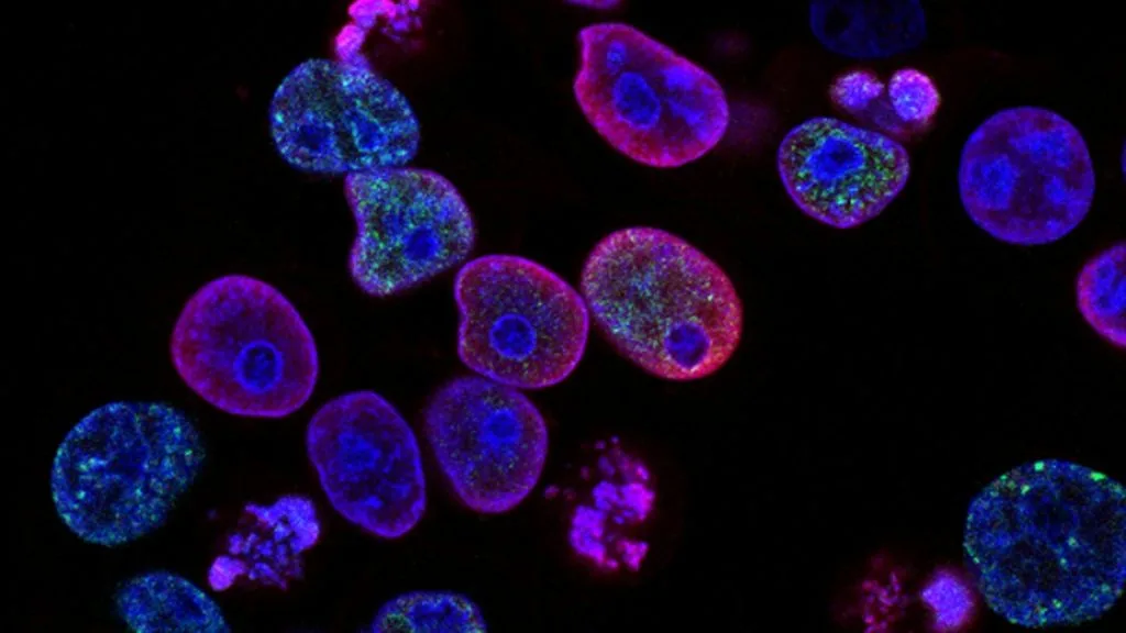
Immune-related response criteria provide standardized radiographic and pathological evaluation models for tracking complex tumor shrinkage dynamics during immune checkpoint blockade. These metrics capture delayed clinical responses and pseudo-progression events driven by immune cell infiltration.
- Radiographic adaptation: Traditional RECIST 1.1 fails to capture pseudo-progression driven by dense **CD8+ cytotoxic T lymphocyte** infiltration.
- Biomarker stratification: Expression of **PD-L1** determined via immunohistochemistry guides front-line monotherapy selection.
- Pathological verification: Post-treatment resections often reveal dense lymphoid aggregates and tertiary lymphoid structures rather than viable carcinoma.
Oncological evaluation of solid tumors treated with **immune checkpoint inhibitors (ICIs)** requires specialized assessment criteria that account for unconventional response patterns. Conventional radiological staging often misclassifies tumor enlargement caused by inflammatory cell influx as true therapeutic failure.
The cellular pathology of regression involves reactivation of exhausted **CD8+ T cells** targeting **PD-1** and **CTLA-4** axes within the tumor microenvironment. Consequently, diagnostic frameworks must integrate pathological tissue evaluation with modified imaging metrics to distinguish active residual disease from treatment-induced necrosis.
CLINICAL & LABORATORY DIAGNOSTIC PROTOCOL
- Specimen acquisition: Biopsy or surgical resection fixed in 10% neutral-buffered formalin (NBF) with cold ischemia time strictly restricted to under 30 minutes.
- Tissue processing: Formalin fixation maintained for 24 to 48 hours at room temperature, followed by paraffin embedding and microtome sectioning at 4-micron thickness onto positively charged adhesive slides.
- Control standards: Tonsil or placental control tissues with validated high and low **PD-L1** expression patterns included in every analytical run to ensure assay reproducibility.
Slide Preparation & Heat-Induced Epitope Retrieval (HIER)
Bake sectioned slides at 60°C for 45 minutes, deparaffinize through three changes of xylene, and rehydrate through descending ethanol grades to distilled water. Perform HIER using a high-pH tris-EDTA buffer (pH 9.0) in a PT Link module maintained at 97°C for 20 minutes.
Peroxidase Blocking & Monoclonal Primary Antibody Incubation
Block endogenous peroxidase activity with 3% hydrogen peroxide solution for 10 minutes at room temperature. Apply clinically validated primary monoclonal antibodies such as clone **22C3** or **SP263** at optimized dilutions and incubate for 30 minutes in a humid chamber.
Polymer Detection & Chromogenic Visualization
Wash slides thoroughly in tris-buffered saline containing 0.05% Tween-20. Apply a horseradish peroxidase-conjugated secondary goat anti-rabbit/mouse polymer reagent for 30 minutes, followed by visualization using a 3,3-diaminobenzidine (**DAB**) chromogen substrate kit for exactly 5 minutes.
Nuclear Counterstaining & Pathological Evaluation
Apply Mayer hematoxylin counterstain for 1 minute, wash in running warm tap water to blue nuclei, dehydrate through ascending ethanols, clear in xylene, and coverslip using permanent mounting medium. Quantitate **tumor proportion score (TPS)** by evaluating membranous staining in a minimum of 100 viable invasive tumor cells.
1. Pathophysiology, Etiology & Cellular Carcinogenesis
Immune checkpoint blockade targets intrinsic regulatory pathways exploited by malignancies to evade immune surveillance. Upregulation of **PD-L1** on tumor cell surfaces engages **PD-1** receptors on tumor-infiltrating lymphocytes, delivering an inhibitory signal that induces T-cell exhaustion and apoptosis.
Mutational burden correlates directly with neoantigen generation, wherein tumors driven by high genomic instability produce aberrant peptides recognizable by major histocompatibility complex molecules. Genetic alterations in DNA mismatch repair machinery (**MSH2**, **MLH1**, **MSH6**, **PMS2**) lead to microsatellite instability and heightened neoantigen loads.
The tumor microenvironment shifts from an immunosuppressive niche characterized by regulatory T cells and myeloid-derived suppressor cells to an active inflammatory field. Successful immunotherapy remodels this stroma, recruiting activated **CD8+ cytotoxic T cells** and natural killer cells capable of eradicating neoplastic clones.
2. Oncogenic Signal Transduction Pathways & Genomic Alterations
Interferon-gamma signaling pathways regulate downstream transcriptional programs that modulate **PD-L1** surface expression on both neoplastic cells and tumor-associated macrophages. Binding of interferon-gamma to its heterodimeric receptor activates receptor-associated Janus kinases **JAK1** and **JAK2**.
Activated kinases phosphorylate signal transducer and activator of transcription 1 (**STAT1**), which translocates to the nucleus to bind the promoter region of the **CD274** gene. Amplifications of chromosome 9p24.1 further drive constitutive **PD-L1** expression independent of cytokine induction.
Oncogenic activation of the **PI3K-AKT-mTOR** pathway in specific carcinoma subtypes upregulates surface **PD-L1** translation through downstream ribosomal protein S6 kinase activation. Targeted inhibition of these pathways combined with checkpoint blockade represents a major strategy to overcome primary resistance.
3. Histopathological Grading, Morphological Subtypes & Differential Diagnosis
Post-immunotherapy tumor specimens frequently display dense lymphocytic aggregates, tertiary lymphoid structures, and extensive granulomatous reactions surrounding degenerating tumor nests. These histological alterations require modified grading systems that differentiate true residual carcinoma from therapy-induced regressive fibrosis.
Immunohistochemical staining panels incorporating **TTF-1**, **Napsin A**, and **p40** remain vital for verifying primary carcinoma histology in treated specimens. Concurrently, multiplex immunohistochemistry allows simultaneous visualization of **CD8**, **CD4**, **FOXP3**, and **PD-L1** to map spatial immune architecture.
Differential diagnosis of treatment-altered tissues includes radiation pneumonitis, opportunistic infections, and sarcoid-like granulomatous reactions. Correlation with clinical histories and serial biomarker assessments ensures accurate pathological interpretation of indeterminate tissue samples.
4. Targeted Therapeutics, Landmark Phase III Trials & Resistance Mechanics
Monoclonal antibodies directed against immune checkpoints, including **pembrolizumab**, **nivolumab**, and **ipilimumab**, have revolutionized systemic oncological management. **Pembrolizumab** binds specifically to **PD-1**, blocking interactions with **PD-L1** and **PD-L2** ligands to restore antitumor T-cell cytotoxicity.
Landmark Phase III clinical trials such as **KEYNOTE-024** and **CheckMate-067** demonstrated superior overall survival and durable progression-free survival advantages for immune checkpoint blockade compared to conventional platinum-based chemotherapy. These trials established **PD-L1 TPS >= 50%** as a definitive predictive biomarker for front-line monotherapy selection.
Mechanisms of acquired resistance include loss of beta-2-microglobulin (**B2M**) compromising antigen presentation, inactivating mutations in **JAK1** or **JAK2**, and upregulation of alternative checkpoint receptors like **LAG-3** and **TIM-3**. Combination regimens involving bispecific antibodies or dual checkpoint inhibition aim to bypass these adaptive resistance pathways.
5. Molecular Staging, Minimal Residual Disease (ctDNA) & Prognostic Stratification
Integration of **AJCC 8th edition** staging parameters with dynamic molecular biomarkers refines risk stratification for patients undergoing immunotherapy. Baseline tumor mutational burden (**TMB**) assessed via next-generation sequencing provides prognostic value independent of histological grade.
Circulating tumor DNA (**ctDNA**) analysis via targeted liquid biopsy assays enables non-invasive tracking of minimal residual disease and early detection of treatment resistance. Clearance of **ctDNA** during early cycles of checkpoint inhibition correlates strongly with prolonged overall survival and durable radiological responses.
Prognostic models now incorporate multiplex immune profiling and serial liquid biopsies to guide duration of therapy and identify candidates for treatment de-escalation. Future clinical paradigms rely on real-time cellular pathology and molecular tracking to personalize immune-oncology interventions.