Instruments Used in Oncology: Cellular Pathology & Histology Analytical Systems, Automated Microtomes, and Molecular Imaging Platforms
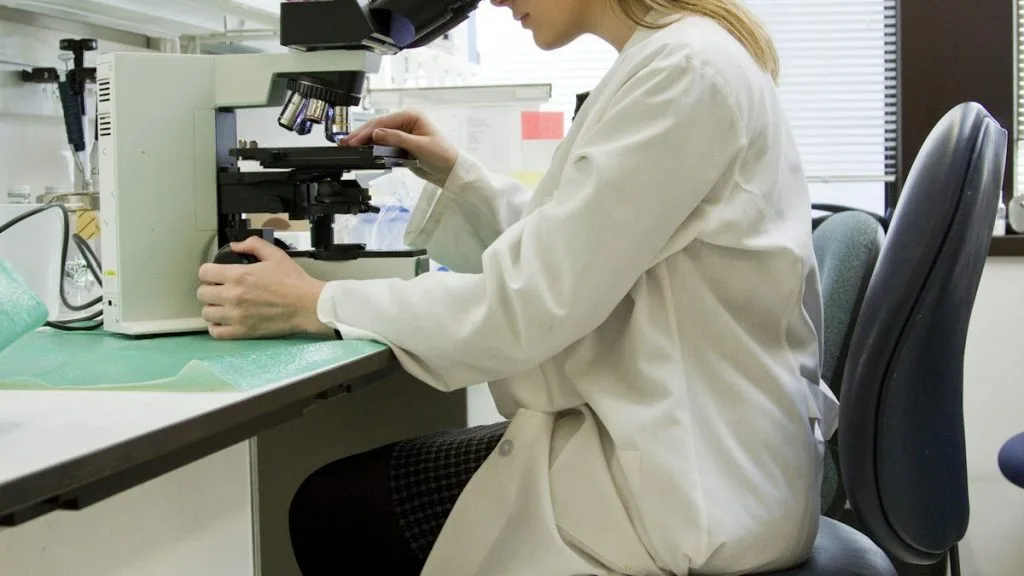
Instruments used in oncology encompass precision clinical laboratory devices, high-throughput automated tissue processors, automated immunohistochemistry stainers, and laser microdissection microscopes. These diagnostic instruments establish definitive cellular pathology, molecular profiling, and spatial phenotyping required for contemporary cancer management.
- Primary analytical infrastructure: Automated vacuum infiltration tissue processors and rotary microtomes producing uniform 4-micron paraffin sections.
- Immunohistochemistry automation: Microprocessor-controlled capillary action stainers ensuring reproducible chromogenic detection of diagnostic oncogenic proteins.
- Molecular pathology integration: Laser capture microdissection and next-generation sequencing sequencers identifying actionable single nucleotide variants.
Cellular pathology and histology laboratories rely on sophisticated mechanical, optical, and digital instruments to process complex oncological specimens. Standardization of tissue fixation, automated embedding, and microtomy directly dictate the analytical validity of downstream genomic assays.
Precision instrumentation minimizes pre-analytical variables, preventing cold ischemia artifact and degradation of nucleic acids or proteins. Modern diagnostic pathology integrates automated staining platforms with digital slide scanners to facilitate high-resolution pathological evaluation.
CLINICAL & LABORATORY DIAGNOSTIC PROTOCOL
- Specimen acquisition: Resected oncological tissue fixed in 10% neutral-buffered formalin (NBF) with a strict cold ischemia time under 60 minutes.
- Instrument calibration: Daily quality control verification of automated microtome blade holders, flotation water baths, and embedding station thermal plates.
- Validation controls: Inclusion of known positive tissue microarrays and negative isotype controls for every automated immunohistochemical run.
Automated Tissue Processing and Vacuum Infiltration
Transfer formalin-fixed tissue cassettes into an automated carousel tissue processor running graded ethanol dehydration, xylene clearing, and molten paraffin wax infiltration under controlled vacuum pressure cycles.
Precision Microtomy and Section Flotation
Mount paraffin blocks on automated rotary microtomes equipped with tungsten carbide blades. Section tissue at a calibrated thickness of 4 microns and float ribbons on a 42°C purified water bath.
Automated Epitope Retrieval and Staining
Load slides onto automated staining instruments (e.g., Ventana Benchmark ULTRA or Dako Omnis) for onboard heat-induced epitope retrieval, blocking, primary antibody incubation, and polymer detection.
Digital Whole Slide Scanning and Pathology Review
Scan finished glass slides using brightfield digital whole slide scanners at 40x magnification with oil immersion optical lenses. Transmit high-resolution digital images to secure pathologist workstations for diagnostic reporting.
1. Tissue Processing, Fixation Instruments, and Vacuum Infiltration Systems
Automated tissue processors utilize microprocessor-controlled fluidics to circulate fixatives, dehydrating alcohols, and clearing agents through porous tissue cassettes. Vacuum infiltration accelerates reagent penetration into dense tumor cores, preserving nuclear morphology for microscopic evaluation.
Precise thermal regulation within paraffin embedding modules prevents tissue scorching and ensures complete block polymerization. Maintenance of rotary microtome blade clearance angles is essential to prevent section compression and chatter artifacts during slicing.
Standardized processing instruments prevent pre-analytical failure modes that obscure malignant cell architecture. Consistency in tissue processing directly supports accurate immunohistochemical staining and subsequent molecular amplification assays.
2. Automated Immunohistochemistry and In Situ Hybridization Platforms
Automated stainers eliminate manual pipetting variability by applying precise volumes of primary antibodies, secondary polymer conjugates, and chromogenic substrates. Instruments such as the Ventana BenchMark ULTRA utilize capillary action and proprietary heating systems to standardize heat-induced epitope retrieval.
Fluorescence in situ hybridization (FISH) automated spotters automate denaturation and hybridization steps for detecting gene amplifications such as **ERBB2** or translocations involving **ALK** and **ROS1**. Automated stringency washes eliminate non-specific probe binding, ensuring crisp fluorescent signal enumeration.
Quality assurance protocols require daily monitoring of instrument fluid lines, nozzle calibrations, and buffer pH levels. Robust instrument maintenance guarantees reproducible staining intensities required for clinical scoring algorithms.
3. Laser Capture Microdissection and Single-Cell Isolation Instrumentation
Laser capture microdissection (LCM) instruments allow pathologists to isolate pure populations of malignant epithelial cells from heterogeneous tumor microenvironments. Ultraviolet or infrared lasers activate specialized transfer films to lift target cells off polyethylene naphthalate membrane slides.
Isolated tumor cells undergo DNA or RNA extraction optimized for low-input next-generation sequencing platforms. This spatial isolation prevents stromal or lymphocytic contamination from confounding variant allele frequency calculations.
Advanced automated microscopes paired with LCM technology incorporate artificial intelligence algorithms to pre-identify malignant glands. Such integration accelerates the isolation of rare cell populations in minimally invasive clinical biopsy specimens.
4. Digital Pathology, Whole Slide Imaging, and Computational Histopathology
Digital whole slide scanners convert physical glass histopathology slides into high-resolution multi-gigabyte digital files using automated robotic stage movements and high-aperture objectives. These imaging platforms facilitate remote telepathology consultations and multi-institutional clinical trial reviews.
Computational pathology software applies machine learning algorithms to quantify biomarker expression, such as calculating tumor proportion scores for **PD-L1** clone 22C3. Automated mitotic index counting and architectural pattern recognition reduce inter-observer variability among pathologists.
Storage infrastructure demands high-capacity server arrays and secure cloud environments compliant with health data privacy regulations. Digital archiving ensures long-term preservation of fragile clinical glass slides without degradation.
5. Molecular Pathology Sequencers and Companion Diagnostic Instrumentation
Next-generation sequencing (NGS) sequencers, including Illumina NovaSeq and Thermo Fisher Ion Torrent systems, decode multiplexed genomic libraries extracted from formalin-fixed paraffin-embedded tissue. These sequencing instruments detect point mutations, insertions, deletions, and copy number variations across panels of oncogenes.
Capillary electrophoresis genetic analyzers perform fragment analysis and Sanger sequencing for validation of complex genomic rearrangements. Mass spectrometry platforms evaluate methylation signatures and specific pharmacogenetic polymorphisms relevant to oncology.
Companion diagnostic instrumentation operates under stringent regulatory frameworks to ensure clinical validity. Precise analytical sensitivity allows detection of low-frequency somatic alterations in circulating tumor DNA liquid biopsies.