Leveraging Microphysiological Systems to Facilitate Neutrophil-Based Cancer Immunotherapy
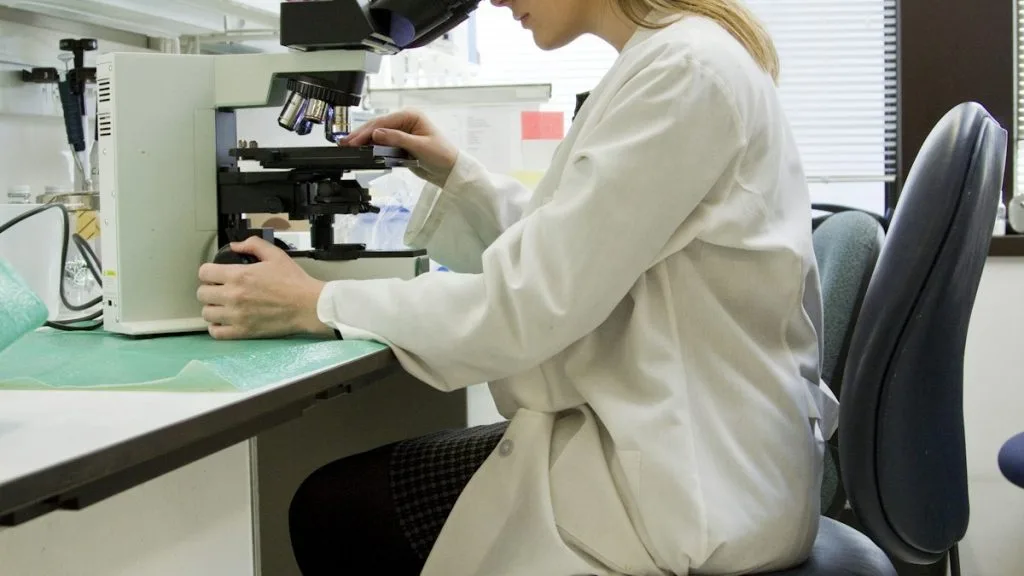
Microphysiological systems (MPS) recreate complex tumor microenvironment niches to optimize the recruitment, activation, and cytotoxic potency of neutrophil-based cancer immunotherapies. These bioengineered platforms bridge the translational gap between in vitro screening and clinical adoptive cellular protocols.
- Microphysiological platforms model complex interstitial gradients, capturing neutrophil chemotaxis via CXCR1 and CXCR2 axis signaling.
- Neutrophil-tumor interactions within microfluidic chambers demonstrate targeted release of neutrophil extracellular traps (NETs) and reactive oxygen species.
- MPS coculture validation enables precise dosing parameters for engineered neutrophil transfer in solid malignancies.
Neutrophils constitute the predominant circulating leukocyte population and increasingly serve as active effector cells in immuno-oncology protocols. Advanced tumor architectures actively secrete interleukin-8 (IL-8) and granulocyte colony-stimulating factor (G-CSF), which drive polymorphonuclear leukocyte infiltration into the stroma.
Conventional 2D culture models fail to replicate the complex vascular perfusion, extracellular matrix density, and mechanical shear stress of the tumor core. Microphysiological systems provide high-throughput, compartmentalized fidelity to dissect neutrophil homing mechanisms and cytotoxicity.
CLINICAL & LABORATORY DIAGNOSTIC PROTOCOL
- Specimen acquisition: Fresh primary tumor resections or engineered 3D bioprinted spheroids processed immediately in sterile phosphate-buffered saline supplemented with 2 percent fetal bovine serum.
- Device fabrication: Polydimethylsiloxane (PDMS) microfluidic chips bonded to glass coverslips via oxygen plasma treatment, sterilized by UV irradiation for 30 minutes.
- Cell isolation: Primary human neutrophils isolated from peripheral blood via negative immunomagnetic selection yielding greater than 98 percent purity and viability.
Microfluidic Chip Seeding & Extracellular Matrix Hydrogel Injection
Inject reconstituted bovine type I collagen or Matrigel matrix mixed with tumor cells into the central channel of the PDMS microphysiological device. Incubate at 37°C for 30 minutes in a humidified 5 percent carbon dioxide incubator to achieve complete hydrogel polymerization.
Vascular Perfusion & Chemokine Gradient Establishment
Connect microfluidic endothelial channels to a calibrated syringe pump perfusion system flowing at a physiological shear stress rate of 2 dyne per square centimeter. Introduce recombinant human IL-8 into the tumor chamber to establish a stable chemotactic gradient across the interstitial matrix.
Neutrophil Introduction & Real-Time Tracking
Introduce CFSE-labeled primary human neutrophils into the vascular inlet channel at a concentration of 1×10^6 cells per milliliter. Utilize confocal laser scanning microscopy equipped with environmental stage controls to record real-time transendothelial migration and interstitial infiltration over 12 hours.
Cytotoxicity Assessment & Multiparameter Immunofluorescence
Fix microfluidic cultures with 4 percent paraformaldehyde for 20 minutes, permeabilize with 0.1 percent Triton X-100, and block with 5 percent normal goat serum. Stain with anti-CD66b, anti-MPO, and cleaved caspase-3 primary antibodies followed by Alexa Fluor secondary conjugates to quantify tumor apoptosis.
1. Pathophysiology, Etiology & Cellular Carcinogenesis
Solid tumors establish systemic inflammatory loops that dynamically reprogram bone marrow myelopoiesis toward immunosuppressive neutrophil phenotypes. Tumor-derived transforming growth factor-beta (TGF-beta) shifts naive neutrophils into tumor-promoting N2 polarizations that facilitate immune evasion.
Epigenetic silencing of pro-inflammatory loci and upregulation of arginase 1 (**ARG1**) and matrix metalloproteinase 9 (**MMP9**) characterize tumor-associated neutrophils. These molecular alterations inhibit local T-cell proliferation and degrade the surrounding extracellular matrix to promote metastasis.
The tumor microenvironment features hypoxic niches that stimulate hypoxia-inducible factor 1-alpha (**HIF-1alpha**), driving chemokine ligand expression. This biochemical signaling network establishes the primary homing tracks that recruit circulating neutrophils directly into the invasive front.
2. Oncogenic Signal Transduction Pathways & Genomic Alterations
Binding of CXCL1, CXCL2, and IL-8 to G protein-coupled receptors **CXCR1** and **CXCR2** initiates intracellular signaling cascades within neutrophils. Receptor engagement activates phospholipase C and triggers intracellular calcium flux essential for directed cellular motility.
Downstream activation of phosphoinositide 3-kinase (**PI3K**) and protein kinase B (**AKT**) pathways regulates actin cytoskeleton remodeling and directional pseudopod protrusion. Concurrently, the MAPK-ERK cascade controls neutrophil survival and degranulation kinetics within the interstitial matrix.
Tumor cells exploit these exact signaling nodes by secreting high levels of myeloid chemoattractants to sequester neutrophils away from secondary lymphoid organs. Pharmacological antagonism of **CXCR2** blocks this recruitment axis, highlighting a key target for microphysiological intervention testing.
3. Histopathological Grading, Morphological Subtypes & Differential Diagnosis
Histological evaluation of neutrophil infiltration relies on immunohistochemical detection of myeloid markers including **CD66b**, **CD15**, and myeloperoxidase (**MPO**). Density scoring divides specimens into neutrophil-poor and neutrophil-enriched infiltrative patterns across advancing tumor margins.
Morphological identification requires differentiating polymorphonuclear neutrophils from tumor-infiltrating lymphocytes and mononuclear macrophages within dense fibrotic stroma. High-resolution microscopy confirms multilobed nuclear architecture and cytoplasmic granule distribution characteristic of functional neutrophils.
Differential diagnostic workflows exclude acute inflammatory abscesses and infection-driven leukocytic infiltrates by correlating tissue histology with negative microbial culture results. Standardized scoring algorithms ensure reproducible quantification of neutrophil density across distinct primary carcinoma subtypes.
4. Targeted Therapeutics, Landmark Phase III Trials & Resistance Mechanics
Translational oncology increasingly investigates adoptive transfer of pre-activated neutrophils engineered via microfluidic priming protocols. Combining these cellular infusions with immune checkpoint inhibitors such as **pembrolizumab** enhances anti-tumor immunity by overcoming local T-cell exclusion.
Landmark clinical investigations such as the KEYNOTE-042 and CheckMate-649 trials underscore the therapeutic necessity of modulating the myeloid compartment in advanced solid tumors. Although direct systemic blockade of neutrophil trafficking has shown limited clinical efficacy, localized microphysiological screening identifies synergy with bispecific antibodies.
Resistance mechanisms within the tumor microenvironment involve neutrophil extracellular trap (**NET**) formation driven by peptidylarginine deiminase 4 (**PAD4**). These web-like chromatin structures shield malignant cells from cytotoxic immune attack and promote intravascular dissemination.
5. Molecular Staging, Minimal Residual Disease (ctDNA) & Prognostic Stratification
Integration of **AJCC 8th edition** tumor staging parameters with systemic inflammatory biomarkers improves prognostic accuracy in gastrointestinal and lung carcinomas. Elevated circulating neutrophil-to-lymphocyte ratios strongly correlate with advanced metastatic burden and inferior progression-free survival.
Liquid biopsy platforms utilizing circulating tumor DNA (**ctDNA**) analysis track clonal evolution alongside dynamic shifts in systemic neutrophil counts. Longitudinal monitoring detects minimal residual disease recurrence well before radiographic imaging manifests clinical relapse.
Prospective clinical paradigms increasingly incorporate microphysiological assay outputs to tailor personalized adoptive immunotherapy schedules. Evaluating patient-specific neutrophil responsiveness ex vivo optimizes therapeutic dosing while minimizing off-target systemic toxicity.