Malignant Neoplasms in Adolescents and Young Adults: Molecular Pathology, Genomic Signatures & Targeted Therapeutics
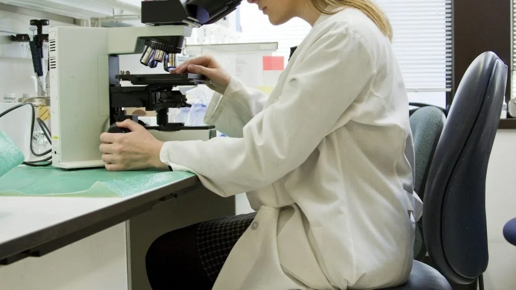
Adolescents and young adults (AYAs), defined as individuals aged 15 to 39 years, experience a unique spectrum of malignancies characterized by distinct embryonal origins and complex genomic rearrangements. Optimal clinical management requires specialized diagnostic protocols and age-adapted therapeutic regimens.
- Primary molecular driver: Chromosomal translocations resulting in chimeric fusion oncoproteins such as EWSR1-FLI1 in Ewing sarcoma.
- First-line standard of care: Multi-agent cytotoxic chemotherapy combined with surgical resection and high-precision radiotherapy.
- Primary mechanism of resistance: Secondary somatic mutations, pathway bypass loops, and enhanced drug efflux transporter expression.
Malignancies in adolescents and young adults (AYAs) exhibit distinct epidemiological and biological features that differ significantly from pediatric and older adult populations. The unique spectrum of diseases includes lymphomas, germ cell tumors, sarcomas, and select carcinomas requiring specialized clinical management.
Oncogenic transformation in this cohort frequently stems from developmental aberrations, epigenetic dysregulation, and characteristic chromosomal translocations. These molecular events drive unchecked cellular proliferation and evasion of apoptosis through specific intracellular signaling pathways.
CLINICAL & LABORATORY DIAGNOSTIC PROTOCOL
- Specimen acquisition: Core needle biopsy or excisional tissue fixed in 10% neutral-buffered formalin (NBF) within 20 minutes of devascularization to preserve nuclear architecture.
- Tissue processing: Fixation duration strictly maintained between 12 and 36 hours at room temperature, followed by paraffin embedding and microtome sectioning at 4 microns.
- Control standards: Internal normal tissue and validated external positive immunohistochemical controls run concurrently with every analytical batch.
Deparaffinization & Heat-Induced Epitope Retrieval (HIER)
Bake slides at 60°C for 45 minutes, pass through three changes of xylene (5 minutes each), and rehydrate through graded ethanol solutions (100%, 95%, 70%) to distilled water. Perform HIER in pH 9.0 EDTA buffer using a pressure cooker or PT module maintained at 97°C for 20 minutes.
Peroxidase Blocking & Primary Antibody Incubation
Block endogenous peroxidase activity by incubating slides in 3% hydrogen peroxide aqueous solution for 10 minutes at room temperature. Apply primary monoclonal antibodies targeting specific diagnostic markers (e.g., **CD99**, **INI1**, or **Myogenin**) at validated titers for 30 minutes.
Polymer Detection & Chromogenic Development
Wash slides thoroughly in Tris-buffered saline with 0.025% Triton X-100. Apply horseradish peroxidase-conjugated secondary polymer reagent for 30 minutes, followed by 3,3-diaminobenzidine (DAB) substrate incubation for 5 minutes.
Nuclear Counterstaining & Permanent Mounting
Counterstain sections with Mayer hematoxylin for 60 seconds, rinse in warm tap water for bluing, dehydrate through graded ethanols, clear in xylene, and apply resinous mounting medium with a glass coverslip. Evaluate staining patterns under brightfield light microscopy.
1. Pathophysiology, Etiology & Cellular Carcinogenesis
AYA malignancies frequently arise from primordial germ cells, mesenchymal stem cells, or lymphoid progenitors arrested during embryogenesis. The cellular transformation is driven by sustained proliferative signaling and evasion of contact inhibition.
Epigenetic dysregulation, including aberrant histone methylation and global DNA hypomethylation, disrupts normal tissue differentiation programs. Key cell cycle checkpoints, including **TP53** and **RB1**, are frequently inactivated via deletion or mutation.
The local tumor microenvironment features an immunosuppressive stroma enriched with cancer-associated fibroblasts and tumor-associated macrophages. These stromal components facilitate angiogenesis and shield malignant cells from immunosurveillance.
2. Oncogenic Signal Transduction Pathways & Genomic Alterations
Ligand-independent receptor tyrosine kinase dimerization initiates potent intracellular cascades across AYA tumors. Structural rearrangements generate constitutively active fusion proteins that bypass physiological regulatory mechanisms.
Downstream signaling proceeds through the **RAS-RAF-MEK-ERK** and **PI3K-AKT-mTOR** pathways to promote cellular survival. Concurrently, transcriptional co-activators stimulate continuous expression of genes required for cell division.
Secondary genomic alterations develop under therapeutic selective pressure, activating alternative bypass tracks. These adaptive changes mediate clinical resistance to conventional cytotoxic and targeted antineoplastic agents.
3. Histopathological Grading, Morphological Subtypes & Differential Diagnosis
Histological evaluation reveals diverse architectural patterns ranging from small round blue cell tumors to pleomorphic sarcomas. Nuclear atypia, frequent mitotic figures, and geographic necrosis characterize high-grade lesions.
Diagnostic immunohistochemical panels incorporate markers such as **CD99** for Ewing sarcoma and loss of **INI1** for rhabdoid tumors. Molecular cytogenetics via fluorescence in situ hybridization confirms pathognomonic translocations.
Differential diagnosis requires ruling out reactive processes, infections, and poorly differentiated carcinomas. Ancillary molecular testing is mandatory to resolve ambiguous morphological phenotypes.
4. Targeted Therapeutics, Landmark Phase III Trials & Resistance Mechanics
Therapeutic strategies combine intensive multi-agent chemotherapy regimens with precision molecular inhibitors directed against specific fusion oncoproteins. Small molecule tyrosine kinase inhibitors target altered receptors like **KIT** and **PDGFRA**.
Landmark clinical trials in AYA oncology, such as those evaluating dose-intensified alkylating regimens, demonstrate improved event-free survival. Incorporation of immunotherapy checkpoint inhibitors continues to reshape treatment paradigms.
Acquired resistance frequently emerges through secondary gatekeeper mutations or activation of parallel survival pathways. Combination therapies and novel antibody-drug conjugates are deployed to overcome refractory disease.
5. Molecular Staging, Minimal Residual Disease (ctDNA) & Prognostic Stratification
Staging algorithms integrate traditional AJCC anatomic parameters with high-throughput genomic profiling data. This combined assessment provides superior prognostic resolution compared to anatomic staging alone.
Circulating tumor DNA (**ctDNA**) analysis via next-generation sequencing enables highly sensitive minimal residual disease detection. Liquid biopsy surveillance detects subclinical recurrence months prior to radiographic progression.
Prognostic stratification models incorporate baseline genomic risk scores, patient age, and treatment response kinetics. These metrics guide individualized post-therapy surveillance and consolidation strategies.