Mass Effect in Intracranial and Solid Organ Malignancies: Cellular Pathology, Biomechanical Compression & Diagnostic Pathophysiology
Mass effect describes the localized tissue displacement, structural distortion, and physiological collapse caused by expanding space-occupying neoplastic lesions. This biomechanical phenomenon drives progressive neurological deficit, parenchymal ischemia, and lethal herniation syndromes across various oncological pathologies.
- Biomechanical stress: Space-occupying neoplasms exert direct hydrostatic and cellular pressure leading to localized vascular collapse.
- Perilesional edema: Upregulation of VEGF via hypoxia-inducible factor 1-alpha drives blood-brain barrier breakdown and vasogenic edema.
- Herniation risk: Unfavorable pressure gradients across intracranial compartments cause subfalcine, transtentorial, or tonsillar herniation.
Mass effect is a critical clinical and pathological manifestation frequently observed in intracranial neoplasms, primary bone tumors, and soft tissue sarcomas. The physical expansion of neoplastic tissue directly compresses adjacent normal parenchyma, inducing regional ischemia and neuroglial dysfunction.
Cellular proliferation outstrips microvascular angiogenesis, creating chronic hypoxia that triggers the secretion of pro-angiogenic factors. This vascular hyperpermeability exacerbates interstitial fluid accumulation, compounding the primary volumetric expansion with extensive secondary edema.
CLINICAL & LABORATORY DIAGNOSTIC PROTOCOL
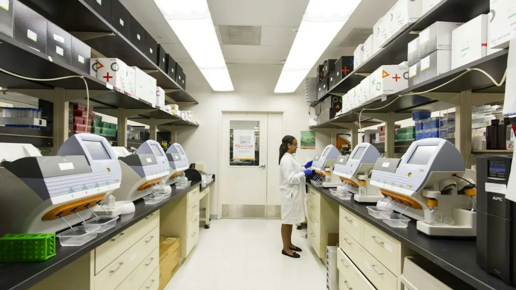
- Specimen acquisition: Stereotactic core needle biopsy or surgical excision fixed in 10% neutral-buffered formalin (NBF) within 15 minutes of devascularization.
- Tissue processing: Formalin fixation maintained strictly for 24 to 48 hours at room temperature; paraffin embedding with 4-micron microtome sectioning on charged adhesion slides.
- Control standards: Internal positive control tissue for glial fibrillary acidic protein (GFAP) and external positive controls for Ki-67 proliferation index run concurrently.
Deparaffinization & Heat-Induced Epitope Retrieval (HIER)
Bake slides at 60°C for 60 minutes, deparaffinize through sequential xylene baths (3 x 5 min), and rehydrate through graded ethanols (100%, 95%, 70%) to deionized water. Perform HIER in target retrieval solution (pH 9.0 EDTA buffer) at 97°C for 20 minutes in a calibrated PT Link module.
Endogenous Peroxidase Block & Primary Antibody Incubation
Block endogenous peroxidase activity with 3% hydrogen peroxide for 10 minutes. Apply optimized primary monoclonal antibody (e.g., anti-GFAP clone GA-5, anti-IDH1 R132H clone H09) at standardized clinical dilution and incubate for 30 minutes at room temperature.
Polymer Detection System & DAB Chromogen Visualization
Wash slides in Tris-buffered saline with 0.05% Tween-20 (TBST). Apply horseradish peroxidase (HRP)-conjugated anti-mouse/rabbit secondary polymer for 30 minutes, followed by incubation with 3,3-diaminobenzidine (DAB) chromogen substrate for 5 minutes.
Counterstaining, Dehydration & Quality Control Scoring
Counterstain with Mayer hematoxylin for 90 seconds, blue in running tap water, dehydrate through graded ethanols, clear in xylene, and apply permanent mounting media. Evaluate staining distribution across viable neoplastic zones under high-power magnification.
1. Pathophysiology, Etiology & Cellular Carcinogenesis
Mass effect originates from unchecked clonal cellular proliferation, driving volumetric expansion within closed anatomical compartments. As the neoplasm enlarges, local compliance thresholds are exceeded, transferring mechanical force to adjacent microvascular networks.
Epigenetic silencing and promoter hypermethylation of tumor suppressor genes such as **PTEN** and **TP53** accelerate dysregulated cell division. This aggressive cellular accumulation transforms the structural architecture of the affected organ parenchyma.
The surrounding microenvironment undergoes significant desmoplasia and hypoxia, activating stromal fibroblasts and tumor-associated macrophages. These cellular alterations release paracrine factors that facilitate matrix remodeling and enhance overall lesion stiffness.
2. Oncogenic Signal Transduction Pathways & Genomic Alterations
Upstream receptor tyrosine kinase activation triggers autophosphorylation cascades that stimulate high-rate cellular replication. Pathways such as the **PI3K-AKT-mTOR** axis drive continuous protein synthesis and evade normal apoptotic signals.
Downstream activation of the **RAS-RAF-MEK-ERK** signaling cascade promotes vascular endothelial growth factor transcription. This molecular signaling directly increases capillary permeability and fuels the aggressive accumulation of perilesional edema.
Genomic instability frequently results in secondary mutations that confer resistance to standard apoptotic stimuli. These intracellular adaptations allow malignant clones to maintain proliferative pressure despite mechanical confinement.
3. Histopathological Grading, Morphological Subtypes & Differential Diagnosis
Histopathological evaluation reveals dense cellular sheets, marked nuclear pleomorphism, and atypical mitotic figures. Growth patterns vary from cohesive lobular structures to highly infiltrative syncytial arrangements invading normal tissue planes.
Diagnostic immunohistochemical panels utilizing **GFAP**, **OLIG2**, and **Ki-67** labeling indices differentiate primary glial neoplasms from metastatic lesions. A **Ki-67** labeling index exceeding 15% correlates strongly with rapid volumetric growth and severe mass effect.
Differential diagnosis requires distinguishing neoplastic mass effect from infectious abscesses, tumefactive demyelinating lesions, and large subacute ischemic strokes. Clinical correlation combined with specialized histochemical stains definitively identifies the underlying etiology.
4. Targeted Therapeutics, Landmark Phase III Trials & Resistance Mechanics
Pharmacological management of mass effect relies on high-dose corticosteroids such as **dexamethasone** to rapidly reduce vasogenic edema by stabilizing tight junctions. Targeted kinase inhibitors and monoclonal antibodies are deployed to halt the underlying proliferative engine.
Landmark clinical trials, including registration studies for anti-angiogenic agents like **bevacizumab**, demonstrate significant reduction in cerebral edema and improvement in progression-free survival. These therapeutic interventions transiently normalize abnormal tumor vasculature and lower intracranial pressure.
Resistance mechanisms frequently emerge through alternative pro-angiogenic pathway activation, such as upregulation of fibroblast growth factor signaling. Combination regimens incorporating immune checkpoint inhibitors are currently evaluated in Phase III protocols to overcome localized treatment resistance.
5. Molecular Staging, Minimal Residual Disease (ctDNA) & Prognostic Stratification
Integration of the **AJCC 8th Edition** TNM staging parameters and WHO grading systems provides critical prognostic stratification. Molecular biomarkers, including **IDH1 R132H** mutations, refine pathological risk models and dictate therapeutic intensity.
Liquid biopsy surveillance utilizing circulating tumor DNA (**ctDNA**) allows non-invasive tracking of minimal residual disease following surgical decompression. Serial quantification of plasma ctDNA variant allele frequencies correlates directly with therapeutic response and recurrence risk.
Overall survival prognostic models incorporate volumetric measurements of mass effect alongside established genomic signatures. These multimodal evaluations optimize patient triage for adjuvant radiation therapy and specialized neuro-oncological care.