MET Exon 14 Skipping Alterations and Capmatinib Efficacy in Non-Small Cell Lung Carcinoma
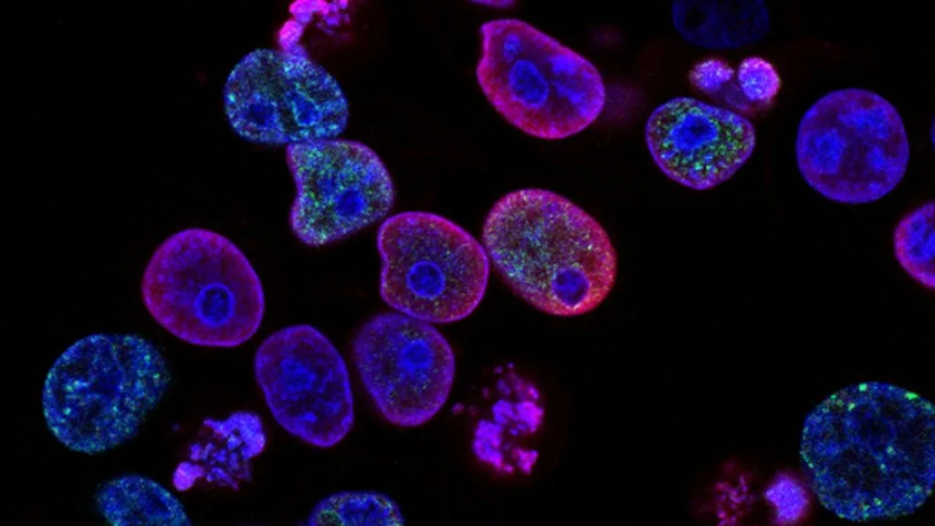
**MET exon 14 skipping mutations** occur in approximately 3% to 4% of lung adenocarcinomas and lead to impaired receptor degradation and aberrant downstream oncogenic signaling. **Capmatinib** is a potent, highly selective oral type Ib **MET receptor tyrosine kinase inhibitor** designed to overcome this oncogenic vulnerability.
- Oncogenic driver mechanism: Splice site mutations involving **MET exon 14** or flanking intronic sequences lead to the deletion of the juxtamembrane domain.
- Protein stability impact: Loss of the **Y1003** binding site for the **CBL E3 ubiquitin ligase** prevents receptor polyubiquitination and subsequent lysosomal degradation.
- Targeted efficacy: **Capmatinib** administration demonstrates robust clinical response rates in treatment-naive and previously treated **MET exon 14-altered** non-small cell lung carcinoma patients.
**MET exon 14 skipping alterations** are frequently identified in older adult populations diagnosed with non-small cell lung carcinoma, demonstrating no absolute predilection for sex and occurring independently of smoking history. These genomic variants encompass diverse point mutations, insertions, or large deletions that disrupt normal pre-mRNA splicing donor and acceptor sites.
The primary functional consequence of **MET exon 14 skipping** is the translation of a truncated **MET receptor** lacking the crucial juxtamembrane domain encoded by exon 14. This structural defect abolishes the negative regulatory feedback loop mediated by **CBL-mediated ubiquitination**, thereby extending the half-life of the activated receptor on the plasma membrane.
CLINICAL & LABORATORY DIAGNOSTIC PROTOCOL
- Specimen acquisition: Tissue core needle biopsy or surgical resection fixed in 10% neutral-buffered formalin (NBF) within 30 minutes of devascularization to maintain cold ischemia time under 1 hour.
- Tissue processing: Formalin fixation time strictly calibrated to 24 to 48 hours at room temperature, followed by paraffin embedding and microtome sectioning at 4-micron thickness onto positively charged adhesive glass slides.
- Control standards: Certified external positive control cell blocks expressing **MET amplification** or known **MET exon 14 skipping variants**, alongside a matched negative isotype control run concurrently for every assay batch.
Slide Deparaffinization & Heat-Induced Epitope Retrieval (HIER)
Bake slides at 60°C for 60 minutes, deparaffinize through sequential xylene baths (3 changes for 5 minutes each), and rehydrate through graded ethanol solutions (100%, 95%, 70%) to deionized water. Perform HIER using a high-pH target retrieval solution containing 10mM Tris base and 1mM EDTA (pH 9.0) at 97°C for 20 minutes in an automated PT Link module.
Endogenous Peroxidase Quenching & Primary Antibody Incubation
Block endogenous peroxidase activity by immersing slides in 3% aqueous hydrogen peroxide for 10 minutes at room temperature, followed by a double rinse in Tris-buffered saline with 0.05% Tween-20 (TBST). Apply optimized primary anti-MET monoclonal antibody (e.g., clone SP44 or D1C2) at a calibrated 1:100 dilution and incubate for 30 minutes in a humidified chamber.
Polymer Detection System & Chromogen Development
Wash slides thoroughly in TBST buffer for 5 minutes. Apply a horseradish peroxidase (HRP)-conjugated goat anti-rabbit polymer secondary reagent and incubate for 30 minutes at room temperature. Visualize immunoreactivity using freshly prepared 3,3-diaminobenzidine (DAB) chromogen substrate solution for exactly 5 minutes.
Nuclear Counterstain, Dehydration & Permanent Mounting
Immerse slides in modified Mayer hematoxylin for 90 seconds for nuclear counterstaining, rinse in running tap water for 3 minutes for bluing, and dehydrate through ascending ethanol series (70%, 95%, 100%). Clear in three changes of xylene and apply a resinous mounting medium beneath a glass coverslip.
1. Pathophysiology, Etiology & Cellular Carcinogenesis
Cellular transformation driven by **MET exon 14 skipping alterations** stems from disruption of canonical donor or acceptor splice sites flanking exon 14 within the **MET** gene locus on chromosome 7q31.2. The resulting aberrant pre-mRNA splicing transcript produces a mutant **MET protein** lacking 47 amino acids encompassing the juxtamembrane regulatory domain.
Epigenetic dysregulation and sustained transcriptional activation subsequently drive constitutive kinase activity without requiring ligand binding by **hepatocyte growth factor (HGF)**. This ligand-independent signaling evades standard intracellular downregulation mechanisms, promoting unchecked cellular proliferation, survival, and epithelial-mesenchymal transition.
The tumor microenvironment in **MET-driven carcinomas** typically exhibits dense vascular networks driven by **MET**-mediated upregulation of **vascular endothelial growth factor (VEGF)**. Concurrently, immune evasion is facilitated via recruitment of myeloid-derived suppressor cells and upregulation of the **PD-1/PD-L1 axis** within the stromal architecture.
2. Oncogenic Signal Transduction Pathways & Genomic Alterations
Ligand binding or structural juxtamembrane deletion induces **MET receptor dimerization** and rapid autophosphorylation of intracellular tyrosine residues (Y1234 and Y1235) within the catalytic loop. These phosphorylated docking sites recruit multiple downstream adapter proteins, initiating multi-pronged oncogenic signal transduction cascades across the malignant cell.
Primary downstream networks include the **RAS-RAF-MEK-ERK pathway** driving transcriptional proliferation and the **PI3K-AKT-mTOR pathway** inhibiting apoptotic cascades and enhancing cellular survival. Concurrently, **STAT3** activation promotes metastatic dissemination, cellular motility, and remodeling of the extracellular matrix.
Secondary resistance mutations within the **MET kinase domain** (such as **D1228** or **Y1230** substitutions) sterically hinder binding of type I **tyrosine kinase inhibitors**, while secondary **MET amplification** bypasses drug-induced pathway inhibition.
3. Histopathological Grading, Morphological Subtypes & Differential Diagnosis
Carcinomas harboring **MET exon 14 skipping alterations** frequently manifest as invasive pulmonary adenocarcinomas with prominent solid, acinar, or papillary architectural growth patterns. Cytological evaluation characteristically reveals significant pleomorphism, large vesicular nuclei, prominent nucleoli, and abundant eosinophilic cytoplasm.
Definitive diagnostic immunohistochemistry requires confirmation of epithelial differentiation via expression of **TTF-1**, **Napsin A**, and **Cytokeratin 7 (CK7)**, while excluding squamous markers such as **p40** and **CK5/6**. **MET overexpression** visualized via immunohistochemistry frequently correlates with underlying genomic alterations but requires orthogonal confirmation.
Differential diagnosis necessitates distinguishing **MET-driven adenocarcinoma** from primary squamous cell carcinoma, large cell neuroendocrine carcinoma, and metastatic malignancies originating from renal cell carcinoma or gastrointestinal tracts.
4. Targeted Therapeutics, Landmark Phase III Trials & Resistance Mechanics
First-generation multi-kinase inhibitors like **crizotinib** demonstrated initial clinical activity against **MET-altered tumors** but possessed limited potency and off-target toxicities. Second-generation selective type I **tyrosine kinase inhibitors**, specifically **capmatinib** and **tepotinib**, were engineered to bind the active conformation of the **MET kinase domain** with high biochemical selectivity.
The pivotal Phase II **GEOMETRY mono-1 trial** evaluated **capmatinib 400mg PO BID** in patients with **MET exon 14-altered non-small cell lung carcinoma**, demonstrating overall response rates exceeding 40% in previously treated cohorts and nearly 70% in treatment-naive cohorts. These landmark findings established **capmatinib** as a standard first-line and salvage therapeutic modality.
Mechanisms of acquired resistance to **capmatinib** involve on-target point mutations at codons **D1228**, **Y1230**, or **F1200**, alongside off-target bypass tracks including **EGFR amplification**, **KRAS mutations**, or **HER2 amplification**.
5. Molecular Staging, Minimal Residual Disease (ctDNA) & Prognostic Stratification
Clinical management requires rigorous integration of the **AJCC 8th edition TNM staging parameters** with comprehensive baseline **next-generation sequencing (NGS)** panels capable of detecting splice site mutations and gene copy number variations. Accurate identification of the **MET exon 14 genotype** directly alters therapeutic decision-making pathways away from standard cytotoxic chemotherapy.
Liquid biopsy surveillance utilizing circulating tumor **DNA (ctDNA)** assays allows for real-time monitoring of minimal residual disease and early detection of emerging secondary **MET resistance mutations** prior to radiologic progression. Quantitative allele frequency tracking provides dynamic prognostic metrics throughout the treatment lifecycle.
Overall survival prognostic stratification remains heavily dependent on baseline performance status, presence of baseline central nervous system metastases, and successful deployment of selective **MET-directed tyrosine kinase inhibitors** upon progression.