Metaplasia in Cellular Pathology: Pathobiological Mechanisms, Epigenetic Reprogramming & Neoplastic Transition
Metaplasia represents the reversible substitution of one fully differentiated adult cell type for another, serving as an adaptive response to chronic environmental injury. Dysregulation of these regenerative pathways frequently transitions into dysplasia, representing a critical cellular bottleneck in epithelial carcinogenesis.
- Primary cellular mechanism: Reprogramming of tissue-specific adult stem cells through microenvironmental cytokine signaling.
- Molecular driver pathways: Aberrant activation of SOX2 and CDX2 transcription factors guiding lineage conversion.
- Malignant transformation: Persistence of chronic injury induces genomic instability, driving progression from metaplasia to invasive carcinoma.
Metaplasia arises predominantly in stratified squamous, glandular, and transitional epithelia subjected to chronic chemical, physical, or infectious irritation. The process involves the replacement of delicate epithelium with more robust cell lineages capable of withstanding hostile microenvironments.
Underlying this histological shift is the direct reprogramming of tissue-specific stem cells and progenitor niches. Pro-inflammatory cytokines and growth factors alter transcriptional networks, shifting cellular differentiation pathways away from the native phenotype.
CLINICAL & LABORATORY DIAGNOSTIC PROTOCOL
- Specimen acquisition: Endoscopic or surgical biopsies fixed in 10% neutral-buffered formalin (NBF) within 15 minutes of devascularization to preserve nuclear architecture.
- Tissue processing: Formalin fixation time calibrated to 18–24 hours at room temperature; paraffin embedding with 3-micron microtome sectioning on charged slides.
- Control standards: On-slide normal tissue controls (such as native squamous mucosa or gastric corpus) run in parallel for every antibody run.
Tissue Deparaffinization & Heat-Induced Epitope Retrieval (HIER)
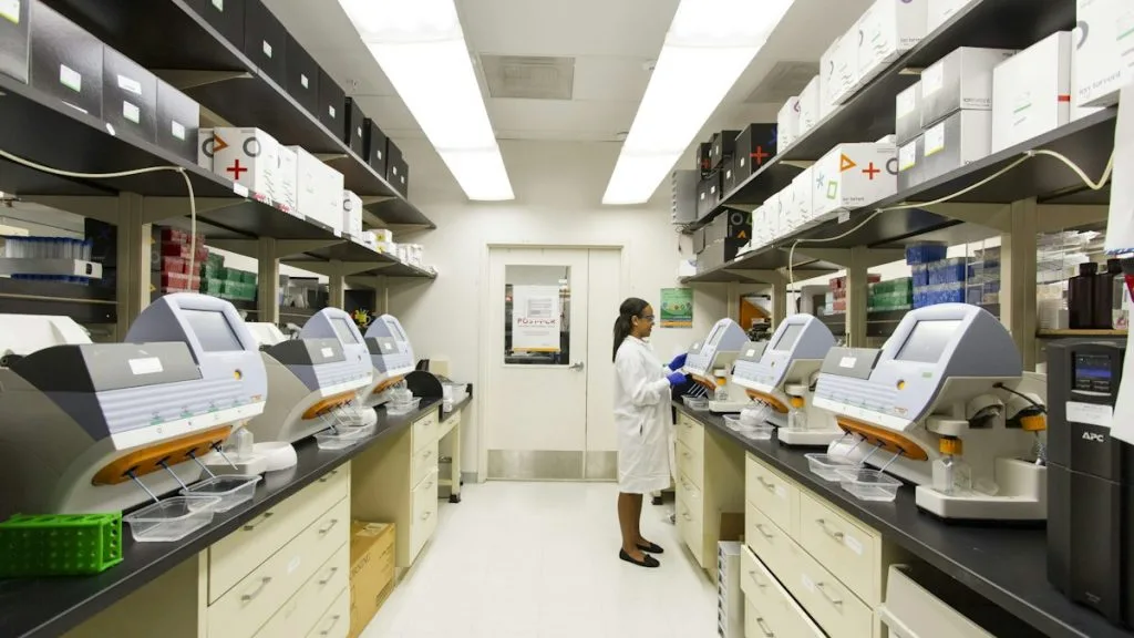
Bake slides at 60°C for 45 minutes, deparaffinize through xylene (3 x 5 min), and rehydrate through graded ethanols (100%, 95%, 70%) to deionized water. Perform HIER in high-pH 9.0 EDTA buffer at 97°C for 25 minutes using an automated retrieval module.
Endogenous Enzyme Quenching & Primary Immunostaining
Block endogenous peroxidase activity with 3% hydrogen peroxide for 10 minutes. Apply primary monoclonal antibodies (e.g., anti-CDX2 clone EPR2764Y or anti-SOX2 clone SP115) at optimized titers and incubate for 32 minutes at room temperature.
Polymer Detection & Chromogenic Visualization
Rinse slides thoroughly in Tris-buffered saline with 0.05% Tween-20 (TBST). Apply horseradish peroxidase-conjugated secondary polymer reagent for 20 minutes, followed by 3,3-diaminobenzidine (DAB) chromogen substrate for 5 minutes.
Counterstaining & Microscopic Evaluation
Apply Mayer hematoxylin counterstain for 90 seconds, blue in mild alkaline tap water, dehydrate through graded ethanols, clear in xylene, and coverslip using permanent mounting medium. Evaluate staining localization in targeted epithelial compartments.
1. Pathophysiology, Etiology & Cellular Carcinogenesis
Metaplasia is driven by chronic physical or chemical stress, such as gastroesophageal reflux inducing Barrett esophagus or tobacco smoke causing respiratory squamous metaplasia. The primary insult induces localized necrosis, prompting robust regenerative signaling from local reserve stem cells.
Epigenetic modifications, including aberrant histone deacetylation and DNA promoter hypermethylation, stably lock progenitor cells into the alternative lineage program. These persistent epigenetic marks create a permissive cellular state for subsequent oncogenic mutational hits.
The surrounding microenvironment features activated cancer-associated fibroblasts, infiltrating macrophages, and secreted interleukins (IL-6, IL-1beta). This inflammatory milieu sustains abnormal proliferation while impairing normal apoptotic DNA damage checkpoints.
2. Oncogenic Signal Transduction Pathways & Genomic Alterations
Metaplastic transformation heavily relies on the dysregulation of homeostatic morphogen cascades, specifically the NOTCH, WNT-beta-catenin, and BMP signaling pathways. Aberrant ligand-receptor engagement drives cellular plasticity and bypasses terminal differentiation arrest.
Downstream activation of the **PI3K-AKT-mTOR** and **RAS-RAF-MEK-ERK** pathways stimulates continuous cellular turnover within the metaplastic epithelium. This heightened mitotic rate dramatically increases the stochastic probability of acquiring secondary driver mutations.
Loss-of-function alterations in **TP53** and amplification events involving **SOX2** or **MYC** are frequently identified in metaplastic fields undergoing dysplastic progression. These molecular aberrations decouple cellular proliferation from normal DNA damage surveillance.
3. Histopathological Grading, Morphological Subtypes & Differential Diagnosis
Histological identification requires recognizing the inappropriate presence of differentiated tissue types within distinct anatomical boundaries, such as gastric or intestinal epithelium within the distal tubular esophagus. Architectural preservation varies, but nuclear polarity and basement membrane integrity remain intact in pure metaplasia.
Diagnostic panels utilize specific immunohistochemical markers, such as **CDX2** and **MUC2** for intestinal metaplasia, or **CK5/6** and **p63** for squamous differentiation. These markers establish lineage fidelity and differentiate true metaplasia from reactive atypia.
The differential diagnosis requires ruling out dysplasia, carcinoma in situ, and well-differentiated adenocarcinoma. The presence of significant pleomorphism, nuclear hyperchromasia, and architectural complexity mandates a reclassification to dysplasia.
4. Targeted Therapeutics, Landmark Phase III Trials & Resistance Mechanics
Management of advanced metaplasia focuses on eliminating pre-neoplastic fields through endoscopic eradication therapies and targeted pharmacological inhibition. Proton pump inhibitors and non-steroidal anti-inflammatory drugs like **celecoxib** mitigate inflammatory stimuli driving progression.
Landmark clinical trials such as the **SURF** trial evaluated endoscopic eradication modalities for Barrett esophagus with high-grade dysplasia, establishing robust progression-free survival benefits. Data from these prospective cohorts confirm that interrupting metaplastic progression reduces esophageal adenocarcinoma incidence.
Refractory metaplastic lesions often evade systemic chemoprevention through alternative receptor tyrosine kinase activation and upregulated multidrug resistance proteins. Combination therapeutic strategies incorporating immune checkpoint inhibitors (**pembrolizumab**) are actively investigated in clinical trials.
5. Molecular Staging, Minimal Residual Disease (ctDNA) & Prognostic Stratification
Staging of metaplastic disorders relies on specialized histological grading systems that quantify the extent and dysplastic grade of the involved mucosal surface. Integrating **AJCC 8th edition** parameters with molecular risk profiling refines clinical surveillance intervals.
Emerging molecular surveillance utilizes circulating tumor DNA (**ctDNA**) and tissue-based next-generation sequencing panels to detect early clonal evolution. Tracking somatic mutations within metaplastic fields provides an objective metric for imminent malignant transformation.
Prognostic stratification categorizes patients based on genomic instability index and epigenetic signatures, guiding personalized surveillance protocols. Prospective validation of these biomarkers continues to optimize clinical management paradigms across oncology networks.