Minimal Residual Disease in Solid Tumors and Hematologic Malignancies: Cellular Pathology, Circulating Tumor DNA, and Clinical Molecular Diagnostics
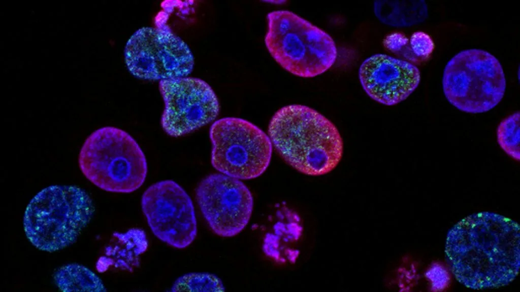
Minimal residual disease refers to the persistence of sub-clinical malignant cells following curative-intent therapy, serving as a primary harbinger of eventual metastatic relapse. Detection utilizes ultra-sensitive molecular diagnostics, including droplet digital PCR, next-generation sequencing, and multiparameter flow cytometry.
- Primary diagnostic utility: Detection of malignant clones at variant allele frequencies (VAF) below 0.01% in peripheral blood or bone marrow.
- Methodological modalities: Tumor-informed ctDNA assays utilizing whole-exome sequencing vs tumor-agnostic methylation panels.
- Clinical implication: Persistence of minimal residual disease identifies patients requiring escalated adjuvant therapy or salvage regimens.
Minimal residual disease assessment represents a paradigm shift in surgical oncology, medical oncology, and clinical pathology. This methodology bridges the chasm between macroscopically clear resection margins and the emergence of macroscopic recurrence.
Molecular pathology laboratories deploy error-corrected sequencing platforms to isolate minute quantities of tumor-derived nucleic acids. The quantification of these circulating genomic fragments permits real-time evaluation of therapeutic efficacy.
CLINICAL & LABORATORY DIAGNOSTIC PROTOCOL
- Specimen acquisition: Peripheral blood collected in Streck Cell-Free DNA BCT tubes (10 mL) or EDTA tubes processed within 2 hours of phlebotomy to prevent genomic DNA contamination from leukocyte lysis.
- Centrifugation protocol: Double-spun plasma extraction protocol consisting of an initial spin at 1,600 x g for 10 minutes followed by high-speed microcentrifugation of supernatant at 16,000 x g for 10 minutes at 4°C.
- Assay validation: Incorporation of synthetic reference standards containing known mutant allele frequencies (0.01% to 1.0%) to monitor limit of detection (LOD) and analytical sensitivity.
Cell-Free DNA Extraction and Quantification
Extract cfDNA from 2 to 4 mL of isolated plasma using automated magnetic bead-based extraction cartridges (e.g., QIAamp Circulating Nucleic Acid Kit). Quantify recovered double-stranded DNA using fluorometric quantitation assays (Qubit dsDNA High Sensitivity Assay Kit).
Tumor-Informed Panel Design and Library Preparation
Sequence archival formalin-fixed paraffin-embedded tumor tissue via targeted NGS panel to identify 16 patient-specific somatic single nucleotide variants (SNVs). Construct multiplex PCR libraries utilizing unique molecular identifiers (UMIs) to eliminate polymerase amplification errors.
Duplex Sequencing and Error Correction
Sequence prepared libraries on high-throughput NGS platforms (Illumina NovaSeq 6000) targeting a minimum raw depth of 100,000x coverage across selected genomic loci. Apply bioinformatic pipelines utilizing UMI consensus building to suppress background sequencing noise.
Variant Analysis and Clinical Reporting
Calculate final variant allele frequency (VAF) and compare against pre-defined analytical validation thresholds. Generate structured pathology report detailing ctDNA status (detected or not detected) alongside quantitative genomic burden.
1. Pathophysiology, Etiology & Cellular Carcinogenesis
Malignant progression involves Darwinian selection of cellular clones capable of evading host immune surveillance and surviving cytotoxic chemotherapy. These therapy-resistant dormant clones often reside within specialized tissue niches, including the bone marrow endosteal space and perineural compartments.
Epigenetic modifications, including aberrant DNA promoter hypermethylation at CpG islands, silence tumor suppressor genes while activating anti-apoptotic signaling networks. These alterations drive sustained cellular proliferation despite profound therapeutic cytotoxicity.
The local tumor microenvironment fosters immune escape by recruiting regulatory T cells, myeloid-derived suppressor cells, and tumor-associated macrophages. These stromal interactions protect occult micrometastases from immune checkpoint inhibition and natural killer cell lysis.
2. Oncogenic Signal Transduction Pathways & Genomic Alterations
Constitutive activation of transmembrane receptor tyrosine kinases triggers downstream signaling cascades that promote survival and proliferation in residual malignant cells. Ligand binding induces receptor homodimerization and intrinsic autophosphorylation of intracellular domain tyrosine residues.
Activated docking sites stimulate the canonical **RAS-RAF-MEK-ERK** pathway to drive transcription of cell cycle progression genes. Simultaneously, the **PI3K-AKT-mTOR** axis is engaged to inhibit apoptosis and upregulate cellular metabolism within dormant clones.
Acquired secondary mutations within kinase domains or parallel bypass tracks enable cancer cells to circumvent targeted pathway inhibition. Persistence of these subclonal genomic alterations dictates clinical progression following apparently curative surgical resection.
3. Histopathological Grading, Morphological Subtypes & Differential Diagnosis
Histopathological examination of primary resection specimens establishes baseline morphological features that correlate with the propensity for micrometastatic dissemination and minimal residual disease persistence. High-grade architectural features, lymphovascular invasion, and perineural infiltration predict elevated post-treatment relapse risks.
Diagnostic panels incorporating lineage-specific immunohistochemical markers, such as **TTF-1**, **Napsin A**, and **CK7** for pulmonary adenocarcinoma, differentiate primary sites from metastatic deposits. Multiparameter flow cytometry panels utilize distinct cluster of differentiation profiles to quantify aberrant cell populations.
Differential diagnosis requires distinguishing residual neoplastic clones from reactive inflammatory infiltrates, therapy-induced stromal atypia, and regenerating benign epithelium. Careful correlation with clinical history and serial molecular assays prevents diagnostic misclassification.
4. Targeted Therapeutics, Landmark Phase III Trials & Resistance Mechanics
Targeted pharmacological interventions, including second- and third-generation tyrosine kinase inhibitors like **osimertinib**, suppress residual oncogenic signaling in molecularly defined populations. Monoclonal antibodies and antibody-drug conjugates deliver cytotoxic payloads directly to cells expressing specific surface antigens.
Landmark Phase III clinical investigations, such as the **ADAURA** trial in **EGFR**-mutated non-small cell lung cancer, demonstrated statistically significant disease-free survival improvements with adjuvant targeted therapy. Similar prospective studies continue to validate ctDNA-guided treatment escalation and de-escalation strategies.
Secondary resistance mechanisms, including tertiary **C797S** mutations and bypass tract amplification, ultimately emerge in patients treated with sustained targeted therapy. Overcoming these resistance clones requires novel bispecific antibodies, combination therapies, and continuous molecular monitoring.
5. Molecular Staging, Minimal Residual Disease (ctDNA) & Prognostic Stratification
Integration of standard **AJCC 8th Edition** TNM staging parameters with high-sensitivity molecular assays refines postoperative risk stratification and clinical management. Conventional radiographic imaging frequently fails to detect microscopic disease burdens below two centimeters in diameter.
Liquid biopsy surveillance using circulating tumor DNA captures real-time dynamic changes in tumor burden during and after adjuvant chemotherapy. Detection of ctDNA following definitive resection correlates with an extremely high probability of clinical relapse.
Prospective incorporation of minimal residual disease status into clinical trial design guides the selective administration of intensive adjuvant therapies to high-risk patients. This precision oncology paradigm minimizes overtreatment for biomarker-negative individuals while optimizing curative potential.