MSI-H / dMMR Deficient Mismatch Repair as a Predictive Agnostic Biomarker: Molecular Pathology, Immunotherapy Response & Clinical Utility
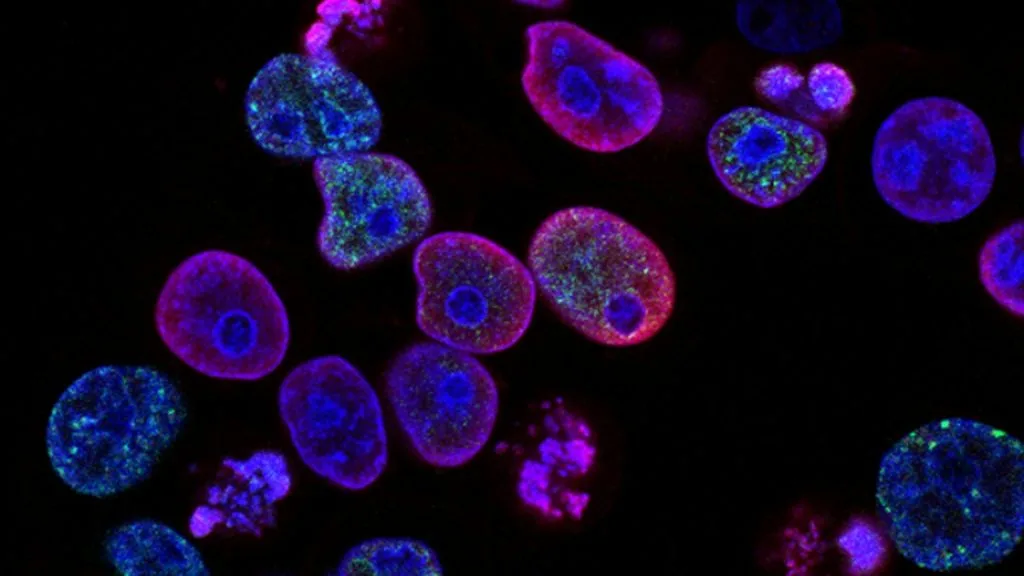
Deficient DNA mismatch repair (dMMR) and high microsatellite instability (MSI-H) constitute a molecular phenotype characterized by hypermutation and neoantigen generation. This validated pan-tumor biomarker predicts high sensitivity to immune checkpoint blockade across multiple solid organ malignancies.
- Primary molecular mechanism: Loss of function in MLH1, MSH2, MSH6, or PMS2 leading to polymerase slippage.
- First-line standard of care: Anti-PD-1 monoclonal antibodies including Pembrolizumab and Nivolumab.
- Diagnostic gold standards: Immunohistochemistry for MMR proteins and multiplex PCR or NGS for microsatellite instability.
Deficient mismatch repair occurs across diverse solid tumors, exhibiting high prevalence in colorectal, endometrial, and gastric adenocarcinomas. The phenotype arises from either germline pathogenic variants defining Lynch syndrome or somatic hypermethylation of the MLH1 promoter.
The resulting DNA replication errors cause extensive length polymorphisms in microsatellite repeat sequences throughout the genome. This hypermutator state generates immunogenic neoantigens that stimulate robust cytotoxic T-cell infiltration within the tumor microenvironment.
CLINICAL & LABORATORY DIAGNOSTIC PROTOCOL
- Specimen acquisition: Formalin-fixed paraffin-embedded (FFPE) tissue blocks or core biopsies fixed in 10% neutral-buffered formalin for 24 to 48 hours.
- Tissue processing: Microtome sectioning at 4-micron thickness onto positively charged glass slides, baked at 60°C for 60 minutes.
- Control standards: Internal normal colonic mucosa, stromal lymphocytes, or endothelial cells acting as positive internal controls for MMR protein expression.
Deparaffinization & Heat-Induced Epitope Retrieval (HIER)
Deparaffinize slides in xylene and rehydrate through descending ethanol concentrations to distilled water. Perform HIER using a high-pH EDTA buffer (pH 9.0) at 97°C for 20 minutes in an automated decloaking chamber.
Peroxidase Blocking & Primary Antibody Application
Block endogenous peroxidase activity with 3% hydrogen peroxide for 10 minutes. Apply optimized monoclonal antibodies targeting **MLH1**, **MSH2**, **MSH6**, and **PMS2** individually, incubating for 30 minutes at room temperature.
Polymer Detection & Chromogenic Visualization
Apply horseradish peroxidase-conjugated secondary polymer reagent for 30 minutes, followed by washing in Tris-buffered saline with Tween-20. Develop signal using 3,3-diaminobenzidine (DAB) chromogen for 5 minutes.
Counterstaining & Pathological Evaluation
Counterstain with Mayer hematoxylin, dehydrate through graded alcohols, clear in xylene, and apply coverslip with permanent mounting medium. Evaluate nuclear staining in invasive tumor cells using internal controls as reference.
1. Pathophysiology, Etiology & Cellular Carcinogenesis
The DNA mismatch repair system corrects base-base mismatches and insertion-deletion loops generated during DNA replication. Inactivation of this pathway via **MLH1**, **MSH2**, **MSH6**, or **PMS2** silencing permits uncorrected replication errors to accumulate.
Epigenetic silencing of **MLH1** through CpG island methylator phenotype (CIMP) represents the predominant etiology in sporadic colorectal carcinomas. Conversely, germline mutations in these repair genes cause Lynch syndrome, an autosomal dominant hereditary cancer predisposition syndrome.
The resulting accumulation of frameshift mutations in coding microsatellites alters reading frames of tumor suppressor genes and regulatory proteins. This molecular cascade drives malignant transformation and accelerates tumorigenesis across diverse tissue lineages.
2. Oncogenic Signal Transduction Pathways & Genomic Alterations
Genomic instability within **MSI-H** tumors targets coding microsatellites located in genes regulating cell cycle progression and apoptosis. Prominent mutational targets include **TGFBR2**, **ACVR2A**, and **BAX**, which sustain unchecked cellular proliferation upon inactivation.
Downstream signaling networks such as the **PI3K-AKT-mTOR** and **MAPK** pathways frequently acquire secondary activating alterations due to the hypermutated genotype. These concurrent alterations shape the evolutionary trajectory and metabolic phenotype of DNA mismatch repair-deficient neoplasms.
The immense mutational burden yields hundreds to thousands of somatic non-synonymous mutations per tumor genome. This proteomic divergence generates novel immunogenic peptides capable of escaping central immune tolerance mechanisms.
3. Histopathological Grading, Morphological Subtypes & Differential Diagnosis
Histologically, **dMMR** adenocarcinomas frequently display distinctive architectural features including medullary growth patterns and prominent intraepithelial lymphocytes. Mucinous differentiation, signet-ring cell components, and peritumoral lymphocytic reactions are also frequently observed.
Immunohistochemical panels evaluating **MLH1**, **MSH2**, **MSH6**, and **PMS2** establish protein loss with high sensitivity and specificity. Aberrant protein profiles guide reflex germline genetic testing to diagnose underlying hereditary cancer syndromes.
Differential diagnosis requires distinguishing primary **dMMR** gastrointestinal or gynecologic carcinomas from metastatic lesions. Diagnostic panels incorporating lineage-specific markers prevent misclassification and direct appropriate clinical staging.
4. Targeted Therapeutics, Landmark Phase III Trials & Resistance Mechanics
Immune checkpoint inhibitors targeting the **PD-1/PD-L1 axis** have revolutionized the management of **MSI-H / dMMR** malignancies. Monoclonal antibodies such as **pembrolizumab** and **nivolumab** reactivate exhausted tumor-infiltrating lymphocytes by blocking inhibitory receptor signaling.
The landmark Phase III **KEYNOTE-177** trial established **pembrolizumab** monotherapy as the standard of care for first-line treatment of metastatic **MSI-H** colorectal cancer. The trial demonstrated a statistically significant improvement in progression-free survival and a favorable safety profile compared to traditional chemotherapy regimens.
Despite high initial response rates, primary and acquired resistance mechanisms to immune checkpoint blockade occasionally emerge. Upregulation of alternative immune checkpoints, beta-2-microglobulin mutations, and defects in antigen presentation machinery drive treatment failure.
5. Molecular Staging, Minimal Residual Disease (ctDNA) & Prognostic Stratification
Integration of **MSI-H** status into AJCC staging paradigms provides critical prognostic stratification for localized disease stages. Stage II **dMMR** colorectal carcinomas demonstrate favorable natural history and lack benefit from adjuvant fluoropyrimidine monotherapy.
Circulating tumor DNA (**ctDNA**) analysis utilizing high-throughput sequencing platforms enables non-invasive detection of minimal residual disease following surgical resection. Dynamic surveillance of **ctDNA** kinetics identifies patients at high risk of disease recurrence before radiographic progression.
Prospective clinical paradigms increasingly utilize agnostic biomarker panels to guide therapeutic escalation and de-escalation strategies. This precision oncology framework optimizes survival outcomes while minimizing treatment-related toxicity across diverse tumor types.