Multipolar Spindles in Cancerous Cellular Pathology: Mitotic Catastrophe, Centrosome Amplification & Genomic Instability
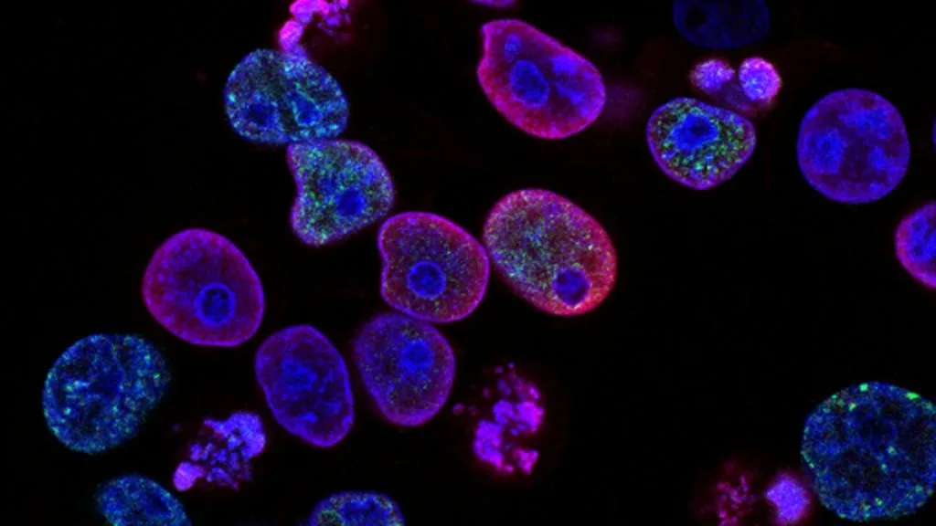
Multipolar spindles represent a severe mitotic aberration characterized by the formation of more than two spindle poles during cell division, driving chromosomal instability and aneuploidy in high-grade human malignancies. Their detection in cellular pathology correlates directly with aggressive tumor behavior, therapeutic resistance, and poor patient survival outcomes.
- Primary cellular mechanism: Centrosome amplification driven by **PLK4** overexpression or cytokinesis failure.
- Pathological consequence: Multipolar mitosis leading to catastrophic chromosomal missegregation and cell death or aneuploidy.
- Therapeutic targeting: Synthetic lethality exploiting mitotic checkpoint vulnerabilities via **AURKA** or **PLK1** inhibition.
Multipolar spindles are frequently observed in high-grade invasive carcinomas, sarcomas, and therapy-refractory tumors exhibiting high mitotic rates. These aberrant mitotic configurations arise predominantly in genetically unstable cells harboring **TP53** inactivating mutations or homologous recombination defects.
The generation of three or more functional spindle poles disrupts normal bipolar chromosome segregation during metaphase to anaphase transition. This cellular chaos results in multi-directional pulling forces, extensive lagging chromosomes, and multipolar anaphase bridges.
CLINICAL & LABORATORY DIAGNOSTIC PROTOCOL
- Specimen acquisition: Fresh surgical resection or core needle biopsy fixed in 10% neutral-buffered formalin (NBF) within 15 minutes of devascularization to preserve delicate mitotic structures.
- Tissue processing: Controlled dehydration and paraffin embedding with 3-micron microtome sectioning on poly-L-lysine coated slides to prevent section detachment during antigen retrieval.
- Control standards: Internal normal proliferative tissues (such as benign tonsil or reactive lymph node germinal centers) utilized to validate basal mitotic spindle staining.
Deparaffinization & High-Temperature Epitope Retrieval
Bake slides at 65°C for 45 minutes, deparaffinize through three changes of xylene (5 minutes each), and rehydrate through descending absolute to 70% ethanols. Perform heat-induced epitope retrieval in a high-pH 9.0 Tris-EDTA buffer using a digital pressure cooker at 125°C for 4 minutes.
Peroxidase Quenching & Centrosome Marker Incubation
Block endogenous peroxidase activity with 3.0% aqueous hydrogen peroxide for 10 minutes at room temperature. Apply primary rabbit monoclonal antibody against **γ-tubulin** (clone EPR15582 at 1:500 dilution) or **pericentrin** and incubate for 60 minutes.
Polymer Amplification & Chromogenic Development
Rinse thoroughly in 0.05% Tween-20 Tris-buffered saline (TBST). Apply anti-rabbit horseradish peroxidase-conjugated polymer secondary reagent for 30 minutes, followed by visualization with 3,3-diaminobenzidine (DAB) chromogen substrate for exactly 7 minutes.
Nuclear Counterstaining & Mitotic Scoring
Counterstain with modified Lillie-Mayer hematoxylin for 90 seconds, blue in alkaline tap water, dehydrate through graded alcohols, clear in xylene, and apply permanent synthetic mounting medium. Quantify multipolar spindle frequency across 50 high-power fields (400x magnification).
1. Pathophysiology, Etiology & Cellular Carcinogenesis
Centrosome amplification is the primary structural precursor to multipolar spindle formation in human solid tumors. Overexpression of master regulatory kinases such as **PLK4** drives excessive centriole duplication during the S-phase of the cell cycle.
Cytokinesis failure and endoreduplication events further compound this anomaly by generating multinucleated giant cells with duplicated centrosome complements. These supernumerary centrosomes frequently cluster to bypass lethal multipolar mitosis, instead forming pseudobipolar spindles that drive viable aneuploidy.
The tumor microenvironment heavily influences spindle integrity through hypoxia-inducible factor signaling, which downregulates bipolarity maintenance factors. Consequently, severe tissue hypoxia directly increases the proportion of multipolar mitotic figures observed in core biopsies.
2. Oncogenic Signal Transduction Pathways & Genomic Alterations
Upstream growth factor receptor activation triggers hyperactive **PI3K-AKT-mTOR** and **RAS-RAF-MEK-ERK** signaling cascades that deregulate cell cycle timing. This sustained proliferative drive overrides the normal dependency on centrosome number for mitotic entry.
Inactivating mutations in tumor suppressor genes such as **TP53** abrogate the post-mitotic checkpoint that normally arrests tetraploid or aneuploid cells. Without functional **p53**, cells possessing multipolar spindles escape apoptosis and propagate unstable genomes.
Defective homologous recombination repair governed by **BRCA1** or **BRCA2** mutations synergizes with centrosome amplification to promote catastrophic chromosomal rearrangements. These genomic aberrations fuel intratumoral heterogeneity and accelerate evolutionary adaptation under therapeutic pressure.
3. Histopathological Grading, Morphological Subtypes & Differential Diagnosis
Histopathological identification of multipolar spindles relies on high-resolution light microscopy examining H&E-stained sections for mitotic figures with three or more distinct chromosomal clusters. These anomalies are predominantly localized in high-grade tumor zones with elevated mitotic indices.
Diagnostic panels incorporating **Ki-67** immunohistochemistry assist in highlighting actively cycling tumor cell populations where multipolar spindles cluster. Special stains are unnecessary, but immunohistochemical mapping of **γ-tubulin** confirms the presence of three or more separated centrosomes.
Differential diagnosis requires distinguishing true multipolar mitoses from multi-nucleated osteoclast-like giant cells or crushing artifacts caused by suboptimal surgical handling. Strict morphological criteria mandate visible chromosomal alignment directed toward three or distinct poles.
4. Targeted Therapeutics, Landmark Phase III Trials & Resistance Mechanics
Pharmacological exploitation of mitotic aberrations focuses on targeting the cellular machinery required for spindle pole clustering. Selective **Aurora kinase A** inhibitors like **alisertib** force cancer cells with amplified centrosomes into lethal multipolar anaphase catastrophes.
Landmark clinical investigations, such as Phase II/III evaluations of **alisertib** in aggressive lymphomas and solid tumors, demonstrate manageable toxicity profiles and localized therapeutic responses. However, acquired resistance frequently emerges through enhanced drug efflux pump expression.
Combination strategies pairing mitotic checkpoint inhibitors with standard cytotoxic chemotherapy agents attempt to overwhelm cancer cell survival thresholds. Despite these efforts, adaptive upregulation of alternative clustering proteins remains a major hurdle in clinical oncology.
5. Molecular Staging, Minimal Residual Disease (ctDNA) & Prognostic Stratification
Integration of multipolar spindle quantification with AJCC 8th edition TNM staging enhances prognostic precision in complex carcinomas. High percentages of multipolar figures consistently correlate with advanced nodal involvement and high histologic grade.
Circulating tumor DNA (**ctDNA**) liquid biopsy surveillance offers dynamic tracking of genomic instability signatures associated with multipolar mitotic rates over time. Serial plasma assays detect emergent subclones before radiologic disease progression becomes evident.
Prognostic stratification models incorporating centrosome amplification metrics allow oncologists to identify patient cohorts requiring intensified adjuvant regimens. Prospective validation of these cellular pathology markers continues to refine personalized clinical management paradigms.