Oncocytic Neoplasia and Cellular Pathology: Mitochondrial Hyperplasia, Genetic Alterations, and Diagnostic Stratification
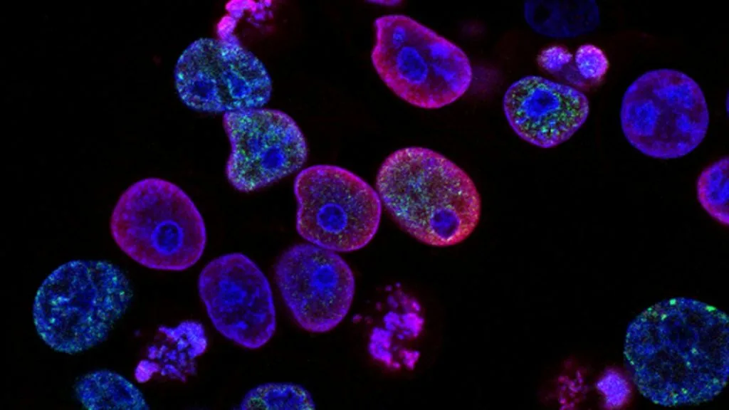
Oncocytic neoplasms are characterized by an abnormal accumulation of dysfunctional mitochondria within epithelial cells, resulting in abundant eosinophilic granular cytoplasm. These tumors arise across multiple organ systems, presenting distinct diagnostic challenges and specific molecular vulnerabilities.
- Primary cellular hallmark: Profuse cytoplasmic accumulation of structurally aberrant, enlarged mitochondria.
- Genetic architecture: Recurrent mutations in mitochondrial DNA complex I genes and nuclear-encoded oxidative phosphorylation regulators.
- Diagnostic requirement: High-power light microscopy confirming abundant granular eosinophilia paired with immunohistochemical exclusion of mimics.
Oncocytic lesions predominantly manifest in endocrine, salivary, and renal tissues among older adult populations with equal gender predilection. These cellular proliferations demonstrate distinct morphological features characterized by uniform cellular enlargement and marked eosinophilic cytoplasmic granularity.
The core pathogenesis involves profound cellular metabolic reprogramming driven by compensatory mitochondrial biogenesis. Defective oxidative phosphorylation complexes trigger perpetual signaling loops that upregulate nuclear respiratory factors to sustain massive mitochondrial accumulation.
CLINICAL & LABORATORY DIAGNOSTIC PROTOCOL
- Specimen acquisition: Fresh surgical resection or core needle biopsy fixed in 10% neutral-buffered formalin (NBF) within 30 minutes of devascularization (cold ischemia time < 1 hr).
- Tissue processing: Formalin fixation time strictly calibrated to 24–48 hours at room temperature; paraffin embedding with 4-micron microtome sectioning on positively charged slides.
- Control standards: On-slide external positive control tissue and validated negative isotype control run in parallel with every analytical batch.
Tissue Deparaffinization & Heat-Induced Epitope Retrieval (HIER)
Bake slides at 60°C for 60 minutes, deparaffinize through sequential xylene baths (3 x 5 min), and rehydrate through graded ethanols (100%, 95%, 70%) to deionized water. Perform HIER in target retrieval solution (pH 9.0 EDTA buffer or pH 6.0 citrate buffer) at 97°C for 20 minutes in a calibrated PT Link module.
Endogenous Peroxidase Block & Primary Antibody Incubation
Quench endogenous peroxidase activity using 3% hydrogen peroxide for 10 minutes. Apply optimized primary monoclonal antibody at calibrated clinical dilution and incubate for 30–60 minutes at room temperature in a humidified chamber.
Polymer Detection System & DAB Chromogen Visualization
Rinse in Tris-buffered saline with 0.05% Tween-20 (TBST). Apply horseradish peroxidase (HRP)-conjugated anti-rabbit/mouse polymer secondary reagent for 30 minutes. Incubate with freshly prepared 3,3-diaminobenzidine (DAB) substrate-chromogen solution for 5-10 minutes until brown precipitates develop.
Counterstaining, Dehydration & Quality Control Scoring
Counterstain nuclei with Mayer hematoxylin for 1–2 minutes, blue in running tap water or 0.08% ammonia water, dehydrate through graded ethanols, clear in xylene, and coverslip with permanent non-aqueous mounting medium. Score tumor cell membrane/nuclear staining under light microscopy using validated scoring algorithm.
1. Pathophysiology, Etiology & Cellular Carcinogenesis
Oncocytic transformation represents a specialized cellular response to profound metabolic stress and impaired adenosine triphosphate generation. Affected epithelial cells undergo massive proliferation of ultrastructural organelles to compensate for enzymatic deficiencies within the electron transport chain.
Epigenetic dysregulation and promoter hypermethylation patterns frequently silence key apoptotic regulators within oncocytic neoplasms. Cell cycle checkpoints become altered, permitting continued division despite sustained mitochondrial genomic instability and elevated reactive oxygen species generation.
The surrounding tumor microenvironment exhibits a dense, hypocellular stroma enriched with cancer-associated fibroblasts and altered extracellular matrix deposition. Hypoxic gradients within larger oncocytomas stimulate neoangiogenesis via sustained autocrine vascular endothelial growth factor signaling pathways.
2. Oncogenic Signal Transduction Pathways & Genomic Alterations
Upstream receptor tyrosine kinase activation initiates downstream signaling cascades that govern mitochondrial biogenesis and cellular hypertrophy. Ligand binding triggers receptor dimerization and autophosphorylation, engaging transcriptional co-activators like peroxisome proliferator-activated receptor gamma coactivator 1-alpha.
Downstream intracellular kinase networks, specifically the **PI3K-AKT-mTOR** axis and the **RAS-RAF-MEK-ERK** pathway, remain constitutively active. This sustained intracellular signaling drives accelerated protein synthesis while simultaneously inhibiting macroautophagy clearance of damaged mitochondria.
Secondary genomic aberrations frequently involve chromosome copy number alterations, large-scale mitochondrial DNA deletions, and loss of heterozygosity at specific tumor suppressor loci. These molecular alterations confer resistance to standard apoptotic stimuli and facilitate local tissue invasion.
3. Histopathological Grading, Morphological Subtypes & Differential Diagnosis
Histopathological evaluation reveals characteristic architectural growth patterns, including solid, trabecular, nested, and microcystic arrangements. Individual cells demonstrate distinct cellular borders, centrally located round nuclei with prominent nucleoli, and deeply eosinophilic granular cytoplasm filled with mitochondria.
Diagnostic immunohistochemical panels utilize markers such as **TTF-1**, **Napsin A**, and **CK7** to pinpoint tissue of origin while excluding non-oncocytic mimics. Chromogenic visualization confirms high mitochondrial enzyme density while establishing baseline immunophenotypic profiles for differential staging.
Differential diagnosis requires strict exclusion of oncogenic hyperplasia, pseudo-oncocytic metaplasia, and metastatic clear cell renal cell carcinoma variants. Meticulous microscopic inspection identifies subtle nuclear inclusions, architectural branching, and capsular or vascular invasion parameters.
4. Targeted Therapeutics, Landmark Phase III Trials & Resistance Mechanics
Systemic management of advanced oncocytic carcinomas traditionally relies on targeted multikinase inhibitors directed against vascular endothelial growth factor receptors and mammalian target of rapamycin pathways. These pharmacologic agents disrupt tumor bioenergetics and suppress uncontrolled cellular proliferation.
Landmark Phase III clinical investigations, including evaluating **mTOR** inhibition protocols, demonstrate modest progression-free survival benefits in refractory oncocytic cohorts. Overall survival endpoints underscore the persistent therapeutic challenge posed by intrinsic metabolic adaptation and drug resistance.
Emerging salvage regimens incorporate novel antibody-drug conjugates and metabolic inhibitors designed to exploit mitochondrial vulnerability profiles. Secondary resistance mechanisms commonly involve bypass activation of alternative survival loops and upregulation of multidrug efflux transporters.
5. Molecular Staging, Minimal Residual Disease (ctDNA) & Prognostic Stratification
Integration of **AJCC 8th edition** TNM staging parameters with high-throughput molecular profiling refines risk stratification across all oncocytic malignancies. Pathologic staging incorporates tumor dimension, angioinvasion status, and extrathyroidal or capsular extension metrics.
Liquid biopsy surveillance employing circulating tumor data (**ctDNA**) tracking enables minimal residual disease detection and early relapse monitoring. Ultra-deep sequencing assays detect somatic mitochondrial and nuclear variants weeks prior to radiographic disease progression.
Prognostic stratification models combine traditional morphological grading with molecular signatures to guide adjuvant therapeutic decision-making. Ongoing clinical trials continue to validate precision oncology paradigms aimed at optimizing patient outcomes through individualized biologic profiling.