Oncogene Addiction in Non-Small Cell Lung Carcinoma: Molecular Pathology, Signal Transduction Networks & Targeted Therapeutics
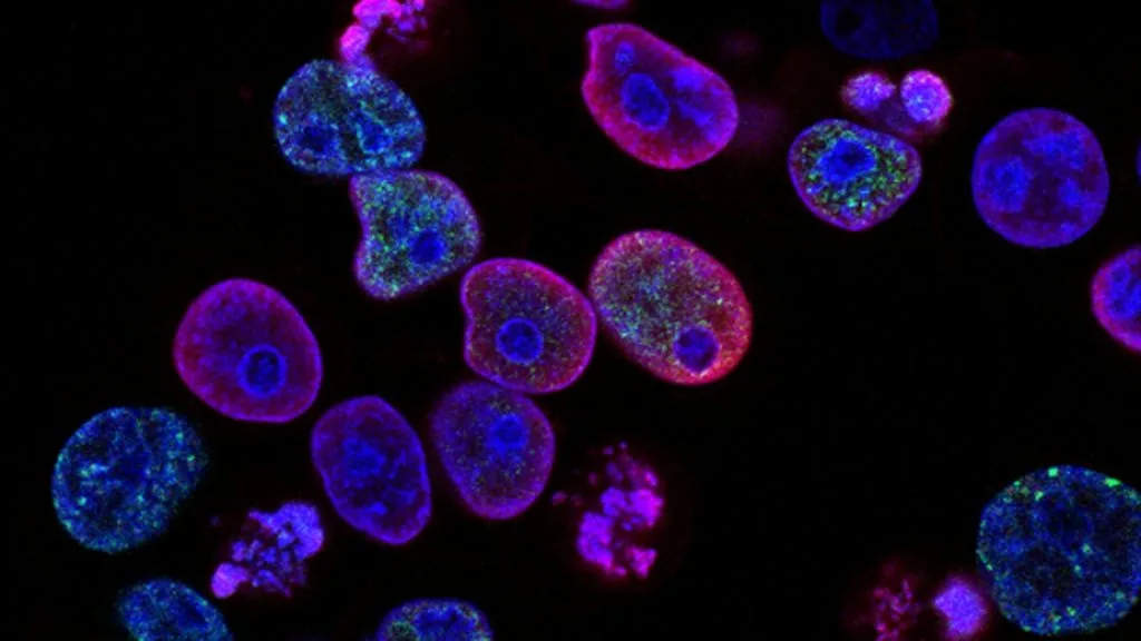
Oncogene addiction defines the absolute dependence of neoplastic cell survival upon a single dominant genomic driver mutation. This biological dependency underpins modern precision oncology and targeted molecular therapeutics.
- Primary molecular driver: Activating mutations or gene translocations in receptor tyrosine kinases such as EGFR and ALK.
- First-line standard of care: Selective third-generation tyrosine kinase inhibitors like osimertinib or alectinib.
- Primary mechanism of resistance: Secondary gatekeeper mutations, bypass pathway activation, or histologic transformation.
Oncogene addiction represents a fundamental paradigm in cellular pathology where tumor cell proliferation and survival rely entirely on a single hyperactivated signaling protein. This molecular vulnerability is frequently observed in lung adenocarcinomas among non-smokers and female patient demographics.
The constitutive activation of driver kinases bypasses normal physiological constraints on cellular growth, apoptosis, and cell cycle progression. Consequently, targeted pharmacologic inhibition of these addicted pathways triggers rapid and profound apoptotic collapse within the tumor parenchyma.
CLINICAL & LABORATORY DIAGNOSTIC PROTOCOL
- Specimen acquisition: Core needle biopsy or surgical resection fixed in 10% neutral-buffered formalin (NBF) within 30 minutes of devascularization with a cold ischemia time strictly under 1 hour.
- Tissue processing: Formalin fixation time calibrated between 24 and 48 hours at room temperature, followed by paraffin embedding and 4-micron microtome sectioning on positively charged slides.
- Control standards: On-slide external positive control tissue and a validated negative isotype control run in parallel with every analytical batch to verify assay specificity.
Tissue Deparaffinization & Heat-Induced Epitope Retrieval (HIER)
Bake slides at 60°C for 60 minutes, deparaffinize through sequential xylene baths (3 x 5 min), and rehydrate through graded ethanols (100%, 95%, 70%) to deionized water. Perform HIER in target retrieval solution using a pH 9.0 EDTA buffer at 97°C for 20 minutes in a calibrated PT Link module.
Endogenous Peroxidase Block & Primary Antibody Incubation
Quench endogenous peroxidase activity using 3% hydrogen peroxide for 10 minutes at room temperature. Apply optimized primary monoclonal antibody clone at a calibrated clinical dilution and incubate for 30 to 60 minutes in a humidified chamber.
Polymer Detection System & DAB Chromogen Visualization
Rinse slides thoroughly in Tris-buffered saline with 0.05% Tween-20 (TBST). Apply horseradish peroxidase-conjugated polymer secondary reagent for 30 minutes and incubate with freshly prepared 3,3-diaminobenzidine substrate-chromogen solution for 5 to 10 minutes.
Counterstaining, Dehydration & Quality Control Scoring
Counterstain nuclei with Mayer hematoxylin for 1 to 2 minutes, blue in running tap water, dehydrate through graded ethanols, clear in xylene, and coverslip with permanent non-aqueous mounting medium. Score tumor cell staining under light microscopy using a validated algorithm.
1. Pathophysiology, Etiology & Cellular Carcinogenesis
Oncogenic transformation begins with somatic DNA damage that disrupts proto-oncogene regulation, converting them into constitutively active drivers. In pulmonary adenocarcinoma, tobacco carcinogens or spontaneous replication errors induce specific point mutations and small in-frame insertions or deletions.
Epigenetic dysregulation often accompanies these genetic events, characterized by aberrant promoter hypermethylation of tumor suppressor loci like CDKN2A and RASSF1A. This methylation silces baseline apoptotic signaling while amplifying proliferative feedback loops.
The tumor microenvironment actively participates in oncogene addiction by assembling an immunosuppressive stroma rich in regulatory T cells and cancer-associated fibroblasts. Aberrant vascularization driven by VEGF overexpression sustains high metabolic demands of the addicted neoplastic clone.
2. Oncogenic Signal Transduction Pathways & Genomic Alterations
Upstream receptor tyrosine kinase activation initiates through ligand-independent homodimerization or heterodimerization, inducing robust autophosphorylation of intracellular tyrosine residues. These phosphorylated docking sites recruit adaptor proteins that trigger intracellular signal propagation.
Downstream signaling diverges primarily through the **RAS-RAF-MEK-ERK** pathway and the **PI3K-AKT-mTOR** axis. These cascades stimulate transcriptional programs that upregulate cyclin D, suppress pro-apoptotic BAD and BAX proteins, and accelerate cellular metabolism.
Secondary gatekeeper mutations, such as **T790M** in **EGFR**, sterically hinder tyrosine kinase inhibitor binding while maintaining adenosine triphosphate affinity. This restoration of kinase activity bypasses drug-induced inhibition and re-establishes oncogenic signaling.
3. Histopathological Grading, Morphological Subtypes & Differential Diagnosis
Histological examination of oncogene-addicted carcinomas reveals distinct architectural patterns including acinar, papillary, solid, and micropapillary arrangements. Cytological features typically display prominent nucleoli, moderate nuclear pleomorphism, and abundant eosinophilic cytoplasm.
A rigorous immunohistochemical panel is mandatory to establish pulmonary lineage, utilizing **TTF-1** positive and **Napsin A** positive staining alongside **CK7**. Squamous markers such as **p40** and **CK5/6** must be absent to confirm adenocarcinoma histology.
The differential diagnosis requires careful exclusion of metastatic malignancies from the breast, gastrointestinal tract, or urothelium. Morphological overlap is resolved through comprehensive clinicopathologic correlation and targeted immunohistochemistry.
4. Targeted Therapeutics, Landmark Phase III Trials & Resistance Mechanics
First- and second-generation reversible tyrosine kinase inhibitors set the baseline for targeted therapy, rapidly superseded by irreversible third-generation agents like **osimertinib**. These molecules selectively target activating mutations while sparing wild-type **EGFR** to minimize cutaneous and gastrointestinal toxicity.
Landmark Phase III trials such as **FLAURA** demonstrated a statistically significant overall survival benefit for frontline **osimertinib** compared to standard EGFR-TKIs. The **ADAURA** trial further established adjuvant **osimertinib** efficacy in resected stage IB-IIIA **EGFR**-mutated non-small cell lung cancer.
Acquired tertiary resistance mutations like **C797S** necessitate novel combination regimens involving fourth-generation inhibitors or antibody-drug conjugates such as **fam-trastuzumab deruxtecan**. Bispecific antibodies like **amivantamab** combined with **lazertinib** effectively target complex MET-amplified escape pathways.
5. Molecular Staging, Minimal Residual Disease (ctDNA) & Prognostic Stratification
Staging integrates the **AJCC 8th edition** TNM classification with multiplex next-generation sequencing assays to establish baseline prognostic categories. Quantitative assessment of allele frequencies provides critical data regarding clonal heterogeneity and tumor burden.
Liquid biopsy surveillance utilizing circulating tumor DNA (**ctDNA**) allows non-invasive detection of minimal residual disease following surgical resection. Dynamic clearance kinetics of plasma **ctDNA** serve as an early surrogate marker for therapeutic efficacy and impending relapse.
Prognostic stratification relies on combining molecular mutation status with residual disease kinetics to guide adjuvant therapy duration. This precision oncology framework optimizes survival outcomes while sparing patients from ineffective systemic cytotoxic chemotherapy.