Oncogenic Cancer Biomarkers in Cellular Pathology: Molecular Signatures, Analytical Validation & Precision Therapeutics
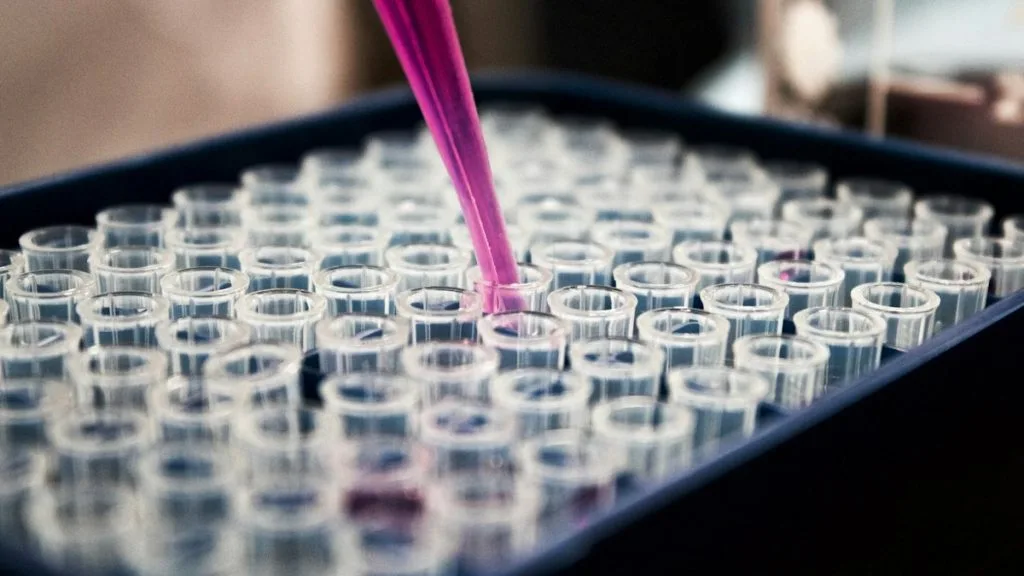
Cancer biomarkers represent quantifiable molecular, cellular, or biochemical alterations that dictate malignant transformation, therapeutic response, and disease surveillance. Comprehensive profiling through next-generation sequencing and immunohistochemistry is mandatory for selecting targeted systemic therapies in oncology.
- Primary molecular alterations include actionable single nucleotide variants, copy number amplifications, and structural chromosomal rearrangements.
- Analytical validation requires strict adherence to pre-analytical tissue handling parameters to preserve nucleic acid and protein integrity.
- Precision oncology relies on companion diagnostics to match molecular subtypes with targeted tyrosine kinase inhibitors and immune checkpoint inhibitors.
Cancer biomarkers span genomic, transcriptomic, proteomic, and epigenomic modalities that drive cellular proliferation and immune evasion across solid tumors. These diagnostic targets dictate therapeutic stratification, directly influencing progression-free survival and overall patient survival metrics.
The integration of multiplex assays allows pathologists to detect low-frequency somatic mutations within heterogeneous tumor microenvironments. Identifying these genomic aberrations is critical for guiding targeted pharmaceutical interventions and monitoring minimal residual disease.
CLINICAL & LABORATORY DIAGNOSTIC PROTOCOL
- Specimen acquisition: Core needle biopsy or surgical resection fixed in 10% neutral-buffered formalin (NBF) within 30 minutes of devascularization with cold ischemia time strictly under 60 minutes.
- Tissue processing: Formalin fixation time calibrated to 24-48 hours at room temperature, followed by paraffin embedding and microtome sectioning at 4-micron thickness onto positively charged adhesive slides.
- Control standards: On-slide external positive control tissue and validated negative isotype control run in parallel with every analytical batch to ensure staining specificity.
Tissue Deparaffinization & Heat-Induced Epitope Retrieval (HIER)
Bake slides at 60°C for 60 minutes, deparaffinize through sequential xylene baths (3 x 5 min), and rehydrate through graded ethanols (100%, 95%, 70%) to deionized water. Perform HIER in target retrieval solution (pH 9.0 EDTA buffer or pH 6.0 citrate buffer) at 97°C for 20 minutes in a calibrated PT Link module.
Endogenous Peroxidase Block & Primary Antibody Incubation
Quench endogenous peroxidase activity using 3% hydrogen peroxide for 10 minutes. Apply optimized primary monoclonal antibody (e.g., clone 22C3, SP263, or 4B5) at calibrated clinical dilution and incubate for 30-60 minutes at room temperature in a humidified chamber.
Polymer Detection System & DAB Chromogen Visualization
Rinse in Tris-buffered saline with 0.05% Tween-20 (TBST). Apply horseradish peroxidase (HRP)-conjugated anti-rabbit/mouse polymer secondary reagent for 30 minutes. Incubate with freshly prepared 3,3-diaminobenzidine (DAB) substrate-chromogen solution for 5-10 minutes until brown precipitates develop.
Counterstaining, Dehydration & Quality Control Scoring
Counterstain nuclei with Mayer hematoxylin for 1-2 minutes, blue in running tap water or 0.08% ammonia water, dehydrate through graded ethanols, clear in xylene, and coverslip with permanent non-aqueous mounting medium. Score tumor cell membrane/nuclear staining under light microscopy using validated scoring algorithm.
1. Pathophysiology, Etiology & Cellular Carcinogenesis
Malignant transformation arises from sequential genomic aberrations that subvert normal cellular homeostasis, apoptosis, and DNA damage repair mechanisms. Somatic mutations accumulate within progenitor stem cells, driving clonal expansion and phenotypic heterogeneity across histological subtypes.
Epigenetic dysregulation, characterized by global DNA hypomethylation and site-specific promoter hypermethylation of tumor suppressor genes, silences critical cell cycle checkpoints. This dysregulation facilitates unchecked cellular division and promotes metastatic dissemination through epithelial-mesenchymal transition.
The tumor microenvironment comprises immunosuppressive cancer-associated fibroblasts, tumor-associated macrophages, and an aberrant extracellular matrix that impedes immune surveillance. Hypoxic signaling pathways stimulate neoangiogenesis via vascular endothelial growth factor upregulation, sustaining metabolic demands of the expanding neoplasm.
2. Oncogenic Signal Transduction Pathways & Genomic Alterations
Upstream receptor tyrosine kinases undergo ligand-dependent or ligand-independent dimerization, inducing conformational shifts and autophosphorylation of intracellular domain tyrosine residues. These phosphorylated motifs serve as docking sites for adaptor proteins, initiating intracellular signal transduction cascades.
Downstream activation of the **RAS-RAF-MEK-ERK** and **PI3K-AKT-mTOR** pathways drives transcriptional programs promoting cellular growth, survival, and metabolic reprogramming. Constitutive activation via activating mutations or gene amplifications bypasses physiological regulatory controls.
Secondary gatekeeper mutations and bypass signaling networks frequently emerge under selective therapeutic pressure from tyrosine kinase inhibitors. These acquired genomic shifts restore downstream survival signaling and necessitate salvage targeted therapies.
3. Histopathological Grading, Morphological Subtypes & Differential Diagnosis
Histopathological evaluation requires assessing architectural growth patterns, such as acinar, papillary, solid, or micropapillary arrangements, alongside nuclear grade and mitotic index. These morphological parameters correlate directly with biologic aggressiveness and metastatic potential.
Diagnostic immunohistochemical panels utilizing specific markers such as **TTF-1**, **Napsin A**, and **CK7** differentiate primary pulmonary adenocarcinomas from squamous cell carcinomas and metastatic lesions. Cross-reactivity is minimized through standardized panels and automated staining platforms.
The differential diagnosis relies on excluding benign reactive proliferations, infectious etiologies, and metastatic neoplasms from distinct primary sites. Correlation with clinical history and high-throughput molecular genotyping resolves ambiguous histological presentations.
4. Targeted Therapeutics, Landmark Phase III Trials & Resistance Mechanics
First-, second-, and third-generation targeted kinase inhibitors selectively bind catalytic domains of mutant proteins, disrupting downstream oncogenic signaling cascades. Small molecule inhibitors and antibody-drug conjugates deliver cytotoxic payloads directly to biomarker-expressing tumor cells.
Landmark Phase III clinical trials, including the **FLAURA** and **ADAURA** trials, demonstrated statistically significant improvements in progression-free survival and overall survival with targeted inhibition compared to standard chemotherapy. These findings established new global standards of care.
Emerging tertiary resistance mutations, such as **C797S**, require novel fourth-generation inhibitors and combination regimens involving bispecific antibodies or antibody-drug conjugates to overcome acquired clinical resistance.
5. Molecular Staging, Minimal Residual Disease (ctDNA) & Prognostic Stratification
Integration of **AJCC 8th Edition** TNM staging parameters with high-resolution molecular biomarker profiles refines prognostic stratification and therapeutic decision-making. Pathological staging quantifies primary tumor invasion, regional lymph node involvement, and distant metastasis.
Liquid biopsy surveillance utilizing circulating tumor **DNA (ctDNA)** enables non-invasive detection of minimal residual disease and early identification of subclinical relapse. Serial monitoring captures dynamic clonal evolution during systemic treatment regimens.
Overall survival prognostic stratification combines clinicopathological variables with multi-omic biomarker assays to guide adjuvant and palliative treatment algorithms in modern oncology practice.