Oncogenic Mutations in Non-Small Cell Lung Cancer: From Molecular Mechanisms to Clinical Practice
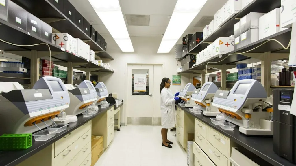
Non-small cell lung carcinoma encompasses a heterogeneous group of malignancies driven by specific genomic alterations requiring precision targeted therapies. Comprehensive molecular profiling guides first-line tyrosine kinase inhibitors and immunotherapy regimens to maximize patient survival.
- Primary molecular driver: In-frame exon 19 deletions or L858R point mutation in ERBB1.
- First-line standard of care: Third-generation irreversible EGFR-TKI (Osimertinib 80mg PO QD).
- Primary mechanism of resistance: T790M gatekeeper mutation or C797S tertiary resistance.
Non-small cell lung cancer accounts for approximately 85 percent of all primary lung malignancies globally. Epidemiological analyses confirm distinct genetic variations between never-smokers carrying targetable driver mutations and heavy tobacco users harboring high tumor mutation burdens.
Cellular transformation is initiated by somatic point mutations, chromosomal translocations, or copy number gains in receptor tyrosine kinases. These structural aberrations constitutively activate downstream survival signaling networks independent of physiological ligand binding.
CLINICAL & LABORATORY DIAGNOSTIC PROTOCOL
- Specimen acquisition: Core needle biopsy or resection fixed in 10% neutral-buffered formalin (NBF) within 30 minutes of devascularization (cold ischemia time < 1 hr).
- Tissue processing: Formalin fixation time strictly calibrated to 24–48 hours at room temperature; paraffin embedding with 4-micron microtome sectioning on positively charged slides.
- Control standards: On-slide external positive control tissue and validated negative isotype control run in parallel with every analytical batch.
Tissue Deparaffinization & Heat-Induced Epitope Retrieval (HIER)
Bake slides at 60°C for 60 minutes, deparaffinize through sequential xylene baths (3 x 5 min), and rehydrate through graded ethanols (100%, 95%, 70%) to deionized water. Perform HIER in target retrieval solution (pH 9.0 EDTA buffer or pH 6.0 citrate buffer) at 97°C for 20 minutes in a calibrated PT Link module.
Endogenous Peroxidase Block & Primary Antibody Incubation
Quench endogenous peroxidase activity using 3% hydrogen peroxide for 10 minutes. Apply optimized primary monoclonal antibody (e.g., clone 22C3, SP263, or 4B5) at calibrated clinical dilution and incubate for 30–60 minutes at room temperature in a humidified chamber.
Polymer Detection System & DAB Chromogen Visualization
Rinse in Tris-buffered saline with 0.05% Tween-20 (TBST). Apply horseradish peroxidase (HRP)-conjugated anti-rabbit/mouse polymer secondary reagent for 30 minutes. Incubate with freshly prepared 3,3-diaminobenzidine (DAB) substrate-chromogen solution for 5-10 minutes until brown precipitates develop.
Counterstaining, Dehydration & Quality Control Scoring
Counterstain nuclei with Mayer hematoxylin for 1–2 minutes, blue in running tap water or 0.08% ammonia water, dehydrate through graded ethanols, clear in xylene, and coverslip with permanent non-aqueous mounting medium. Score tumor cell membrane/nuclear staining under light microscopy using validated scoring algorithm.
1. Pathophysiology, Etiology & Cellular Carcinogenesis
Carcinogenesis in non-small cell lung cancer arises from cumulative genetic and epigenetic insults to alveolar type II pneumocytes or bronchial epithelial stem cells. Environmental carcinogens induce characteristic transversion mutations, whereas adenocarcinomas in non-smokers frequently harbor spontaneous transition errors.
Epigenetic dysregulation involving promoter hypermethylation of tumor suppressor genes such as **CDKN2A** and **RASSF1A** silences critical cell cycle checkpoints. This molecular silencing permits unchecked cellular proliferation and evasion of apoptosis within the pulmonary parenchyma.
The surrounding tumor microenvironment exhibits significant cellular plasticity characterized by immunosuppressive regulatory T cells, tumor-associated macrophages, and dense fibrotic stroma. Neovascularization is driven by sustained autocrine upregulation of vascular endothelial growth factor within hypoxic niches.
2. Oncogenic Signal Transduction Pathways & Genomic Alterations
Upstream receptor tyrosine kinase activation involves ligand-independent dimerization and auto-phosphorylation of intracellular domain tyrosine residues. Structural rearrangements, such as **EML4-ALK** and **KIF5B-RET** fusions, generate constitutively active chimeric proteins.
Activated receptors propagate intracellular signals via the **RAS-RAF-MEK-ERK** mitogen-activated protein kinase cascade and the **PI3K-AKT-mTOR** survival pathway. These signaling networks drive relentless transcriptional activation of genes regulating cell cycle progression and metabolic reprogramming.
Acquired resistance frequently emerges through secondary gatekeeper mutations like **EGFR T790M** or activation of bypass tracks such as **MET** amplification. These biochemical alterations restore downstream signaling despite effective inhibition of the primary oncogenic target.
3. Histopathological Grading, Morphological Subtypes & Differential Diagnosis
Adenocarcinomas exhibit diverse architectural growth patterns categorized into acinar, papillary, solid, and micropapillary subtypes. High-grade components correlate directly with aggressive clinical behavior and increased risk of vascular and pleural invasion.
A rigorous diagnostic immunohistochemical panel requires nuclear positivity for **TTF-1** and cytoplasmic staining for **Napsin A** to confirm pulmonary adenocarcinoma. Squamous cell carcinomas are distinguished by positive expression of **p40**, **p63**, and cytokeratin 5/6.
Differential diagnosis requires ruling out metastatic extrapulmonary malignancies and benign reactive processes through correlation with clinical history and specialized immunohistochemical markers. Exact tumor cell percentage must be documented for valid molecular profiling.
4. Targeted Therapeutics, Landmark Phase III Trials & Resistance Mechanics
First- and second-generation tyrosine kinase inhibitors successfully bound **EGFR** reversibly but were limited by acquired resistance and off-target toxicities. Third-generation irreversible inhibitors like **osimertinib** selectively target both sensitizing mutations and the **T790M** resistance mutation while sparing wild-type receptors.
The landmark **FLAURA** Phase III clinical trial demonstrated a statistically significant overall survival benefit for first-line **osimertinib** compared to older generation tyrosine kinase inhibitors. Similarly, the **ADAURA** trial established practice-changing adjuvant **osimertinib** efficacy in resected stage IB-IIIA **EGFR**-mutant cases.
Tertiary resistance mutations such as **C797S** necessitate fourth-generation inhibitors and combinatorial antibody-drug conjugates like **fam-trastuzumab deruxtecan** for **HER2**-overexpressing tumors. Bispecific antibodies such as **amivantamab** combined with **lazertinib** provide robust response rates in refractory settings.
5. Molecular Staging, Minimal Residual Disease (ctDNA) & Prognostic Stratification
Staging integration requires combining **AJCC 8th edition** anatomic parameters with multiplex genomic profiling to determine precise risk stratification. The presence of actionable drivers alters therapeutic decision-making independent of standard TNM classification.
Liquid biopsy surveillance utilizing circulating tumor data derived from cell-free DNA enables early detection of minimal residual disease post-resection. Quantitative droplet digital polymerase chain reaction and targeted next-generation sequencing track variant allele frequencies over time.
Prognostic stratification models incorporate molecular clearance kinetics, PD-L1 tumor proportion scores, and baseline genomic tumor burden to predict recurrence. These precision oncology frameworks drive individualized surveillance and tailored adjuvant treatment algorithms.