Pathophysiological Mechanisms of Cancer Pain: Cellular Pathology, Perineural Invasion, and Nociceptive Transduction
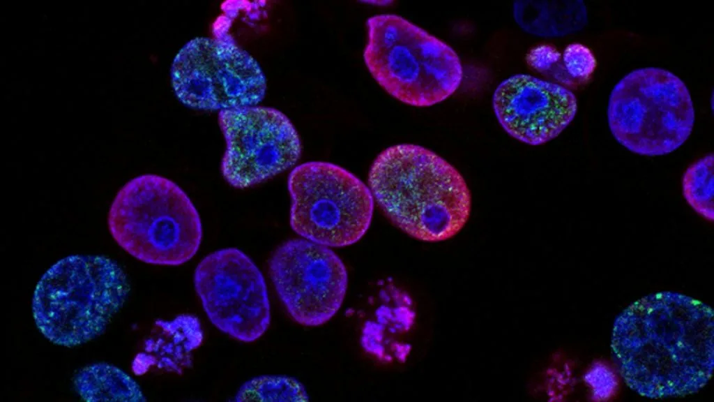
Cancer pain arises from complex cellular interactions, direct tissue destruction, and neuroimmune dysregulation within the tumor microenvironment. Understanding these nociceptive mechanisms enables targeted pharmacotherapy and specialized interventional oncology management.
- Primary nociceptive driver: Upregulation of neurotrophic factors such as Nerve Growth Factor (**NGF**) and Brain-Derived Neurotrophic Factor (**BDNF**).
- Structural neural destruction: Perineural invasion driven by **S100**-positive Schwann cell remodeling and axonal degeneration.
- Bone destruction pathway: **RANKL**-mediated osteoclast activation driving severe skeletal related events and breakthrough pain.
Cancer-related pain impacts over seventy percent of patients with advanced metastatic malignancies, presenting profound cellular pathology challenges. The etiology stems from a combination of direct somatic nerve compression, visceral distension, and inflammatory cytokine release.
Uncontrolled tumor growth forces mechanical distortion of neighboring stromal compartments and activates peripheral nociceptors. This process triggers sustained action potentials along primary afferent C-fibres and A-delta fibers directly into the dorsal horn.
CLINICAL & LABORATORY DIAGNOSTIC PROTOCOL
- Specimen acquisition: Surgical resection or core biopsy fixed in 10% neutral-buffered formalin (**NBF**) for 24 to 48 hours to preserve neural architecture.
- Tissue processing: Paraffin embedding followed by 4-micron microtome sectioning onto positively charged adhesive glass slides.
- Control standards: Peripheral nerve segments and dorsal root ganglia utilized as positive internal controls for neurofilament and S100 immunohistochemistry.
Deparaffinization & Heat-Induced Epitope Retrieval
Bake slides at 60°C for 60 minutes, pass through xylene (3 x 5 min), and rehydrate through descending alcohols to distilled water. Perform antigen retrieval in pH 9.0 Tris-EDTA buffer at 97°C for 20 minutes inside an automated pressure chamber.
Endogenous Blockade & Primary Antibody Incubation
Block endogenous peroxidases with 3% hydrogen peroxide for 10 minutes, followed by protein block. Apply primary monoclonal antibody targeting **S100** or **neurofilament** at validated clinical dilutions for 45 minutes at room temperature.
Polymer Conjugation & Chromogenic Visualization
Apply horseradish peroxidase-conjugated secondary polymer for 30 minutes at room temperature. Wash thoroughly in Tris-buffered saline with Tween-20 and develop using 3,3-diaminobenzidine (**DAB**) for exactly 5 minutes.
Counterstaining & Microscopic Perineural Assessment
Counterstain nuclei with Harris hematoxylin for 60 seconds, dehydrate through graded ethanols, clear in xylene, and apply permanent mounting media. Evaluate sections under light microscopy to quantify malignant cell infiltration within neural sheaths.
1. Pathophysiology, Etiology & Cellular Carcinogenesis
Malignant cellular proliferation directly compresses adjacent somatic and autonomic neural networks. This structural impingement induces localized ischemia, hypoxia, and subsequent localized metabolic acidosis within the extracellular matrix.
Tumor cells secrete various proteolytic enzymes, including matrix metalloproteinases (**MMPs**), degrading the perineurium and endoneurium. This degradation exposes sensory nerve endings to direct biochemical irritation from tumor metabolites.
The local microenvironment becomes densely infiltrated by tumor-associated macrophages (**TAMs**) and neutrophils. These immune cells release pro-inflammatory cytokines such as **IL-1beta**, **IL-6**, and **TNF-alpha**, directly sensitizing peripheral nociceptors.
2. Oncogenic Signal Transduction Pathways & Genomic Alterations
Oncogenic activation frequently upregulates neurotrophic factors including **NGF** and its high-affinity tyrosine kinase receptor **TrkA**. Binding of **NGF** to **TrkA** on sensory nerve terminals initiates retrograde axonal transport and central sensitization.
Downstream intracellular signaling via the **PI3K-AKT-mTOR** and **RAS-RAF-MEK-ERK** pathways amplifies expression of transient receptor potential vanilloid 1 (**TRPV1**) channels. Upregulation of **TRPV1** lowers the activation threshold of nociceptors, producing thermal and mechanical hyperalgesia.
Autocrine and paracrine loops involving endothelin-1 (**ET-1**) acting on **ET_A** receptors further stimulate nociceptive firing. This autocrine loop correlates directly with the intensity of bone and visceral breakthrough pain.
3. Histopathological Grading, Morphological Subtypes & Differential Diagnosis
Perineural invasion (**PNI**) is defined histologically as tumor tracking along or encircling nerve bundles through more than thirty-three percent of the circumference. This morphological feature is graded semiquantitatively based on nerve diameter and depth of infiltration.
Immunohistochemical staining panels utilizing **S100** and smooth muscle actin (**SMA**) assist in delineating nerve fibers from adjacent fibrovascular tissue. **CD56** and synaptophysin highlight neuroendocrine differentiation commonly associated with neuropathic pain syndromes.
Differential diagnosis requires distinguishing true malignant perineural invasion from benign pseudoinvasion or treatment-induced radiation fibrosis. Careful morphological analysis prevents staging errors and misattribution of pain etiologies.
4. Targeted Therapeutics, Landmark Phase III Trials & Resistance Mechanics
Analgesic management relies on precise WHO analgesic ladder protocols combined with targeted biologic therapies. Bisphosphonates and **RANKL** inhibitors such as denosumab directly neutralize osteoclast activity to mitigate bone destruction pain.
Landmark clinical investigations demonstrate that monoclonal antibodies directed against **NGF**, such as tanezumab, significantly reduce osteoarthritic and metastatic bone pain. Phase III trials highlight potent analgesic efficacy while necessitating close monitoring for sympathetic joint complications.
Refractory cancer pain driven by persistent neuroplastic remodeling often requires intrathecal drug delivery systems. Continuous infusion of ziconotide, a selective **N-type voltage-sensitive calcium channel blocker**, targets dorsal horn nociceptive transmission directly.
5. Molecular Staging, Minimal Residual Disease (ctDNA) & Prognostic Stratification
Integration of **AJCC 8th edition** TNM parameters with pain severity scores provides a comprehensive prognostic index for advanced disease. High baseline pain scores frequently correlate with advanced nodal burden and visceral metastasis.
Circulating tumor DNA (**ctDNA**) analysis allows non-invasive tracking of clonal evolution and metastatic progression before radiographic manifestation. Detecting rising **ctDNA** allele frequencies alerts clinicians to expanding tumor volume driving new pain sites.
Multimodal prognostic stratification models now incorporate molecular biomarkers of neuro-invasion alongside traditional clinicopathologic staging. This quantitative approach guides aggressive palliative interventions and improves overall quality of life.