Recent Trends of Incidence, Mortality, Treatment, and Overall Survival of Hepatocellular Carcinoma in the United States
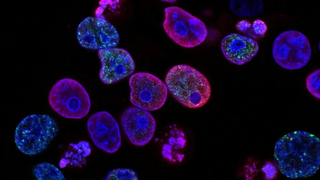
Hepatocellular carcinoma represents the predominant primary malignancy of the liver with rising incidence trends across the United States. Contemporary management integrates multidisciplinary staging with advanced systemic immunotherapies and targeted molecular tyrosine kinase inhibitors to improve overall survival.
- Incidence dynamics: Shifting demographic profiles driven by metabolic dysfunction-associated steatotic liver disease (MASLD) alongside viral hepatitis.
- Frontline standard of care: Combination immunotherapy utilizing **atezolizumab** plus **bevacizumab** or **durvalumab** plus **tremelimumab**.
- Resistance and progression: Second-line therapeutic switching to multikinase inhibitors **sorafenib**, **lenvatinib**, **regorafenib**, or **cabozantinib**.
Hepatocellular carcinoma incidence in the United States has undergone significant epidemiological shifts over the past two decades. While chronic hepatitis C virus infections have historically dominated etiology, metabolic syndrome and MASLD now drive a parallel rise in non-viral carcinogenesis.
Malignant transformation stems from chronic hepatic necroinflammation, oxidative stress, and fibrogenesis progressing to cirrhosis. This pathological microenvironment induces continuous hepatocyte regeneration, genomic instability, and activation of proliferative oncogenic signaling pathways.
CLINICAL & LABORATORY DIAGNOSTIC PROTOCOL
- Specimen acquisition: Ultrasound-guided core needle biopsy of hepatic lesions fixed in 10% neutral-buffered formalin (NBF) within 20 minutes of devascularization.
- Tissue processing: Formalin fixation time calibrated to 24–48 hours at room temperature; paraffin embedding followed by 4-micron microtome sectioning onto charged slides.
- Control standards: On-slide external positive control tissue and validated negative isotype control run in parallel with every immunohistochemical batch.
Tissue Deparaffinization & Heat-Induced Epitope Retrieval
Bake slides at 60°C for 60 minutes, deparaffinize through sequential xylene baths (3 x 5 min), and rehydrate through graded ethanols (100%, 95%, 70%) to deionized water. Perform heat-induced epitope retrieval in target retrieval solution (pH 9.0 EDTA buffer) at 97°C for 20 minutes in a calibrated automated module.
Endogenous Peroxidase Block & Primary Antibody Incubation
Quench endogenous peroxidase activity using 3% hydrogen peroxide for 10 minutes. Apply optimized primary monoclonal antibody (e.g., anti-Glypican-3 clone 1G12, anti-Arginase-1, or anti-PD-L1 clone 22C3) at calibrated clinical dilution and incubate for 30 minutes at room temperature.
Polymer Detection System & DAB Chromogen Visualization
Rinse in Tris-buffered saline with 0.05% Tween-20. Apply horseradish peroxidase-conjugated anti-rabbit/mouse polymer secondary reagent for 30 minutes. Incubate with freshly prepared 3,3-diaminobenzidine substrate-chromogen solution for 5-10 minutes until brown precipitates develop.
Counterstaining, Dehydration & Quality Control Scoring
Counterstain nuclei with Mayer hematoxylin for 1-2 minutes, blue in running tap water, dehydrate through graded ethanols, clear in xylene, and coverslip with permanent mounting medium. Score tumor cell membrane and cytoplasmic staining under light microscopy using validated diagnostic criteria.
1. Pathophysiology, Etiology & Cellular Carcinogenesis
Hepatocellular carcinoma development is intimately linked to chronic hepatic injury, inflammation, and cirrhosis. Etiological factors include chronic hepatitis B virus integration, hepatitis C infection, alcohol-related liver disease, and MASLD.
Epigenetic dysregulation, frequent **TERT** promoter mutations, and aberrant DNA methylation silence tumor suppressor loci while activating proliferative cascades. Progressive telomere shortening in hepatocytes drives chromosomal instability and cellular transformation.
The tumor microenvironment features an immunosuppressive stroma enriched with regulatory T cells and myeloid-derived suppressor cells. Hepatic stellate cells differentiate into cancer-associated fibroblasts, secreting extracellular matrix proteins that promote angiogenesis and metastasis.
2. Oncogenic Signal Transduction Pathways & Genomic Alterations
Upstream receptor tyrosine kinases, including **VEGFR**, **PDGFR**, and **EGFR**, become aberrantly activated through autocrine and paracrine ligand loops. Ligand binding triggers receptor dimerization and intracellular domain autophosphorylation.
Downstream signaling diverges into the **RAS-RAF-MEK-ERK** cascade and the **PI3K-AKT-mTOR** pathway, driving cellular proliferation and inhibiting apoptosis. Concurrently, activating **CTNNB1** mutations drive Wnt-beta-catenin signaling abnormalities.
Genomic profiling identifies frequent inactivating mutations in **TP53** and **ARID1A**, which disrupt DNA damage repair and chromatin remodeling. These alterations contribute to therapeutic resistance and aggressive phenotypic progression.
3. Histopathological Grading, Morphological Subtypes & Differential Diagnosis
Histological evaluation reveals trabecular, pseudoglandular, or solid architectural growth patterns with varying degrees of cytological atypia. Nuclear pleomorphism, multinucleated giant cells, and bile production confirm hepatocellular differentiation.
Diagnostic immunohistochemical panels utilize **Arginase-1**, **Glypican-3**, and **HepPar-1** to distinguish hepatocellular carcinoma from intrahepatic cholangiocarcinoma. **CD34** highlights aberrant sinusoidal capillarization.
Differential diagnosis requires ruling out metastatic adenocarcinomas and neuroendocrine tumors through comprehensive immunohistochemical staining profiles. Reticulin staining helps evaluate loss of normal reticulin framework in dysplastic nodules.
4. Targeted Therapeutics, Landmark Phase III Trials & Resistance Mechanics
First-line systemic therapy has evolved from single-agent multikinase inhibitors to immune checkpoint blockade combinations. The combination of **atezolizumab** targeting **PD-L1** and **bevacizumab** targeting **VEGF** established a new overall survival benchmark.
The IMbrave150 clinical trial demonstrated superior overall survival and progression-free survival with **atezolizumab** plus **bevacizumab** compared to **sorafenib**. Similarly, the HIMALAYA trial established **durvalumab** plus **tremelimumab** as a validated frontline immunotherapeutic regimen.
Secondary resistance mechanisms involve activation of alternative angiogenic pathways and compensatory signaling loops. Salvage therapies such as **regorafenib**, **cabozantinib**, and **lenvatinib** target refractory progression pathways.
5. Molecular Staging, Minimal Residual Disease (ctDNA) & Prognostic Stratification
Staging integrates traditional **AJCC 8th Edition TNM** parameters with functional liver reserve metrics including the Child-Pugh classification. The Barcelona Clinic Liver Cancer system guides treatment allocation based on tumor burden and performance status.
Emerging surveillance modalities utilize circulating tumor DNA (**ctDNA**) assays to detect minimal residual disease following surgical resection or ablation. Longitudinal liquid biopsy tracking identifies molecular recurrence prior to radiographic detection.
Prognostic stratification relies on integrating clinical variables, baseline alpha-fetoprotein levels, and genomic signatures. This precision oncology framework optimizes therapeutic sequencing and improves long-term survival outcomes.