ROS1 Proto-Oncogene Fusions in Pulmonary Adenocarcinoma: Molecular Pathology, Clinical Diagnostic Paradigms & Tyrosine Kinase Therapeutics
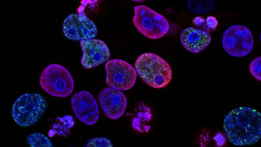
ROS1 proto-oncogene rearrangements drive approximately 1 to 2 percent of advanced pulmonary adenocarcinomas, representing a distinct molecular subset characterized by younger patient age and never-smoking status. Targeted tyrosine kinase inhibitor therapy yields high objective response rates, establishing mandatory multi-modality molecular profiling at initial diagnosis.
- Primary molecular driver: Chromosomal translocations involving the 6q22 locus of ROS1 fused to diverse fusion partners.
- First-line standard of care: Selective small-molecule tyrosine kinase inhibitors including crizotinib, entrectinib, and repotrectinib.
- Primary mechanism of resistance: Acquired secondary kinase domain mutations such as the solvent-front G2032R substitution.
Pulmonary adenocarcinoma harboring **ROS1** fusions typically manifests in younger non-smoking patients without significant tobacco exposure history. This genomic aberration constitutes approximately 1 to 2 percent of non-small cell lung cancer cases globally.
The oncogenic mechanism relies on constitutive activation of the **ROS1** receptor tyrosine kinase catalytic domain via oligomerization domains contributed by fusion partners. This constitutive kinase signaling drives unchecked proliferation through downstream mitogenic cascades.
CLINICAL & LABORATORY DIAGNOSTIC PROTOCOL
- Specimen acquisition: Formalin-fixed paraffin-embedded core needle biopsy or surgical resection specimen fixed in 10% neutral-buffered formalin for 6 to 72 hours.
- Tissue processing: Microtome sectioning at 4-micron thickness mounted on positively charged glass slides with optimal drying at 60 degrees Celsius.
- Control standards: Positive control lung adenocarcinoma tissue with known **ROS1** rearrangement and negative isotype control processed in parallel.
Deparaffinization & Heat-Induced Epitope Retrieval
Bake slides at 60°C for 60 minutes, pass through three changes of xylene (5 minutes each), and rehydrate through graded ethanols (100%, 95%, 70%) to distilled water. Perform heat-induced epitope retrieval in a high-pH EDTA buffer (pH 9.0) at 97°C for 20 minutes using an automated staining module.
Endogenous Peroxidase Block & Primary Antibody Application
Inubate sections in 3% hydrogen peroxide for 10 minutes to quench endogenous tissue peroxidase activity, followed by rinsing in Tris-buffered saline with 0.05% Tween-20. Apply anti-**ROS1** rabbit monoclonal primary antibody (clone D4D6) at a 1:100 dilution and incubate for 30 minutes at room temperature.
Polymer Detection & Chromogen Development
Apply horseradish peroxidase-conjugated anti-rabbit secondary polymer reagent for 30 minutes at room temperature. Visualize immunoreactivity by applying 3,3-diaminobenzidine substrate-chromogen solution for 10 minutes, generating a stable brown cytoplasmic precipitate.
Counterstaining, Dehydration & Interpretation
Counterstain nuclei with Mayer hematoxylin for 60 seconds, wash in warm tap water for bluing, dehydrate through graded ethanols, clear in xylene, and coverslip using a non-aqueous permanent mounting medium. Evaluate granular cytoplasmic staining in tumor cells and confirm positivity against internal endothelial controls.
1. Pathophysiology, Etiology & Cellular Carcinogenesis
Pulmonary adenocarcinomas with **ROS1** fusions arise from transformed alveolar type II pneumocytes or terminal respiratory bronchiolar epithelial cells. These alterations occur independently of tobacco carcinogen exposure signatures, showing no enrichment for transition or transversion mutational burdens.
The molecular pathogenesis centers on balanced or unbalanced chromosomal translocations resulting in the juxtaposition of the **ROS1** kinase domain to the 5-prime partner gene. This structural rearrangement removes auto-inhibitory regulatory domains and forces constitutive tyrosine phosphorylation.
The resulting tumor microenvironment typically exhibits an immunosuppressive phenotype with variable infiltration by regulatory T lymphocytes and myeloid-derived suppressor cells. Tumor vascularity is promoted through autocrine vascular endothelial growth factor upregulation driven by downstream transcriptional effectors.
2. Oncogenic Signal Transduction Pathways & Genomic Alterations
Ligand-independent dimerization of the chimeric **ROS1** fusion protein initiates autophosphorylation of critical intracellular tyrosine residues. These phosphorylated motifs act as docking sites for Src homology 2 domain-containing adaptor proteins.
Activated docking complexes stimulate the classical **RAS-RAF-MEK-ERK** pathway, promoting accelerated cell cycle progression through cyclin D1 induction. Concurrently, the **PI3K-AKT-mTOR** axis is triggered, inhibiting apoptotic pathways and sustaining high metabolic rates via aerobic glycolysis.
Secondary resistance mechanisms emerge via acquired mutations within the **ROS1** kinase domain, most notably the **G2032R** solvent-front substitution that sterically hinders drug binding. Other bypass tracks involve parallel amplification of **MET**, **KRAS** codon mutations, or histologic transformation to small cell lung carcinoma.
3. Histopathological Grading, Morphological Subtypes & Differential Diagnosis
Histologically, **ROS1**-rearranged adenocarcinomas frequently display solid, acinar, cribriform, or papillary growth architectures accompanied by abundant signet-ring cell morphology. Intracellular mucin production and psammoma bodies are recognized diagnostic clues observed during routine hematoxylin and eosin evaluation.
Diagnostic immunohistochemical panels demonstrate robust co-expression of **TTF-1** and **Napsin A**, confirming primary pulmonary adenocarcinoma lineage. Squamous markers such as **p40** and **CK5/6** remain consistently negative, excluding squamous cell carcinoma.
Differential diagnosis requires ruling out metastatic mucinous adenocarcinomas originating from the gastrointestinal tract or ovary. Comprehensive clinical history, imaging correlation, and pulmonary-specific immunohistochemical markers resolve ambiguity in challenging diagnostic biopsies.
4. Targeted Therapeutics, Landmark Phase III Trials & Resistance Mechanics
First-generation multi-targeted tyrosine kinase inhibitors like **crizotinib** established initial targeted paradigms by inhibiting **ROS1**, **ALK**, and **MET**. Second- and third-generation agents such as **entrectinib**, **repotrectinib**, and **lorlatinib** deliver superior central nervous system penetration and potency against resistance variants.
The landmark PROFILE 1001 Phase I/II trial demonstrated an objective response rate of 72 percent and a median progression-free survival of 19.2 months with **crizotinib**. Subsequent pivotal data from the **STRATA** and **TRIDENT-1** trials confirmed exceptional intracranial efficacy and prolonged progression-free survival for next-generation macrocyclic inhibitors like **repotrectinib**.
Acquired tertiary resistance driven by **G2032R** or **L2196M** mutations necessitates next-generation sequencing surveillance at progression. Salvage strategies employ next-generation ROS1 inhibitors, covalent inhibitors, or combination trials incorporating immune checkpoint inhibitors.
5. Molecular Staging, Minimal Residual Disease (ctDNA) & Prognostic Stratification
Accurate staging incorporates the **AJCC 8th Edition TNM** classification system alongside mandatory baseline multiplex molecular profiling. Defining metastatic burden and baseline central nervous system involvement dictates initial systemic therapy sequencing versus consolidative stereotactic radiation.
Liquid biopsy surveillance utilizing circulating tumor DNA (**ctDNA**) via targeted next-generation sequencing enables minimal residual disease detection and early identification of emergent resistance alleles. Longitudinal **ctDNA** monitoring correlates closely with radiographic progression and guides timely therapeutic switching.
Prognostic stratification in advanced disease depends on central nervous system control, performance status, and the specific structural fusion partner. Patients receiving genotype-matched targeted therapies experience significantly extended overall survival exceeding 50 months in clinical cohorts.