Single-Cell Transcriptomic Analysis of Tumor Heterogeneity and the Microenvironment in Pseudomyxoma Peritonei
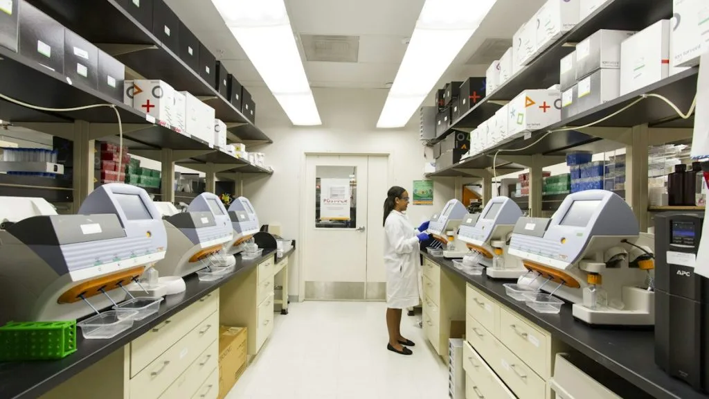
Pseudomyxoma peritonei is a rare neoplastic condition characterized by gelatinous ascites and peritoneal dissemination, predominantly originating from appendiceal mucinous neoplasms. Single-cell transcriptomic analyses reveal profound cellular heterogeneity within both the epithelial tumor compartment and the immunosuppressive tumor microenvironment.
- Single-cell RNA sequencing reveals distinct epithelial subclones harboring concurrent KRAS and GNAS mutations driving mucin hypersecretion.
- The tumor microenvironment exhibits a profoundly immunosuppressive phenotype dominated by exhausted CD8+ T cells and M2-polarized tumor-associated macrophages.
- Cancer-associated fibroblasts (CAFs) actively remodel the extracellular matrix through upregulation of collagen and fibronectin transcripts.
Pseudomyxoma peritonei affects approximately one to two per million individuals annually, demonstrating a slight female predominance secondary to ovarian involvement termed Krukenberg-like mucinous tumors. The clinical presentation is frequently insidious, often diagnosed incidentally during evaluation for abdominal distension or acute appendicitis.
Oncogenic activation in pseudomyxoma peritonei is driven by persistent missense mutations in the **KRAS** and **GNAS** genes. These molecular events constitutively stimulate downstream signaling cascades including **MAPK/ERK** and **cAMP/PKA**, resulting in unchecked cellular proliferation and copious mucin glycoprotein production.
CLINICAL & LABORATORY DIAGNOSTIC PROTOCOL
- Specimen acquisition: Fresh peritoneal mucinous tumor tissue or cellular ascites obtained immediately during cytoreductive surgery with cold ischemia time strictly restricted under 20 minutes.
- Cell dissociation: Enzymatic digestion utilizing collagenase type IV (2 mg/mL) and DNase I (100 U/mL) in RPMI-1640 medium at 37°C for 45 minutes with continuous gentle agitation.
- Single-cell suspension: Filtration through a 40-micron cell strainer, red blood cell lysis using ammonium-chloride-potassium buffer, and viability assessment via trypan blue exclusion ensuring greater than 85 percent live cells.
Droplet-Based Single-Cell RNA Sequencing Library Construction
Load viable single-cell suspensions into a Chromium Controller (10x Genomics) targeting a recovery of 6,000 to 10,000 cells per capture channel. Perform reverse transcription inside oil droplets to generate complementary DNA tagged with cellular barcodes and unique molecular identifiers (UMIs).
High-Throughput Next-Generation Sequencing
Sequence amplified cDNA libraries on an Illumina NovaSeq 6000 platform utilizing a paired-end dual-index sequencing format. Target a minimum sequencing depth of 50,000 reads per cell to achieve adequate transcriptomic coverage of lowly expressed genes.
Bioinformatic Quality Control and Alignment
Align raw sequencing reads to the human reference genome (GRCh38) utilizing Cell Ranger software v7.0. Filter out low-quality cells exhibiting fewer than 500 detected genes, greater than 25,000 UMIs, or mitochondrial gene expression exceeding 20 percent of total cellular transcripts.
Dimensionality Reduction and Cell Type Annotation
Execute principal component analysis (PCA) for dimensionality reduction followed by Uniform Manifold Approximation and Projection (UMAP) visualization. Annotate distinct cellular clusters using canonical lineage markers including **MUC2** for goblet-like epithelial cells, **PTPRC** for immune cells, and **COL1A1** for fibroblasts.
1. Pathophysiology, Etiology & Cellular Carcinogenesis
Pseudomyxoma peritonei originates predominantly from perforated low-grade appendiceal mucinous neoplasms. The primary appendiceal lesion breaches the muscularis propria, disseminating neoplastic epithelial cells into the peritoneal cavity where they establish persistent mucin-producing implants.
Epigenetic dysregulation characterized by aberrant promoter hypermethylation silences critical DNA mismatch repair genes and tumor suppressors. This epigenetic shift maintains the neoplastic cells in a perpetually proliferative yet apoptosis-resistant state within the peritoneal cavity.
The peritoneal tumor microenvironment is defined by an acellular mucin pool that shields epithelial tumor nests from host immune surveillance. Extensive desmoplasia driven by activated stromal cells creates physical barriers that impede systemic drug delivery and immune cell infiltration.
2. Oncogenic Signal Transduction Pathways & Genomic Alterations
Transmembrane receptor activation initiates downstream intracellular signaling cascades that govern mucin secretion and cellular proliferation in pseudomyxoma peritonei. Activating missense mutations in **KRAS** at codon 12 lock the protein in an active GTP-bound conformation.
Constitutively active **KRAS** perpetually drives the **RAF-MEK-ERK** kinase cascade while parallel **PI3K-AKT-mTOR** pathway activation inhibits apoptotic signaling. Concurrently, **GNAS** mutations stimulate adenylate cyclase, elevating intracellular cyclic adenosine monophosphate and driving hypersecretion of **MUC2** and **MUC5AC** mucins.
Secondary genomic alterations involving **TP53** loss or **SMAD4** inactivation correlate with histological progression from low-grade to high-grade peritoneal mucinous carcinomatosis. These advanced genetic aberrations bypass standard cell cycle checkpoints and accelerate invasive clinical phenotypes.
3. Histopathological Grading, Morphological Subtypes & Differential Diagnosis
Histopathological classification categorizes pseudomyxoma peritonei into low-grade appendiceal mucinous carcinoma (LAMN), high-grade appendiceal mucinous carcinoma (HAMN), and mucinous adenocarcinoma with signet ring cells. Low-grade lesions exhibit bland epithelial stratification with minimal nuclear atypia embedded in abundant extracellular mucin.
Diagnostic immunohistochemical panels are mandatory to confirm appendiceal origin and exclude primary ovarian or colonic adenocarcinomas. The standard panel demonstrates diffuse positivity for **CK20**, **CDX2**, **MUC2**, and **MUC5AC**, coupled with diagnostic negativity for **CK7**.
Differential diagnosis requires distinguishing pseudomyxoma peritonei from peritoneal mesothelioma and metastatic pancreaticobiliary mucinous carcinomas. Careful morphologic evaluation combined with immunohistochemical staining for mesothelial markers like **WT1** and **calretinin** resolves ambiguous diagnostic dilemmas.
4. Targeted Therapeutics, Landmark Phase III Trials & Resistance Mechanics
Conventional cytotoxic chemotherapy regimens historically demonstrated limited efficacy in pseudomyxoma peritonei due to the hypovascular nature of peritoneal implants. Therapeutic paradigms rely heavily on complete cytoreductive surgery combined with hyperthermic intraperitoneal chemotherapy using **mitomycin C** or **oxaliplatin**.
Landmark surgical oncology trials published by Sugarbaker and colleagues established cytoreductive surgery and HIPEC as the standard of care, demonstrating five-year overall survival rates exceeding 80 percent for low-grade disease. Emerging clinical trials evaluate targeted inhibition of downstream **MEK** and **mTOR** pathways in refractory **KRAS**-mutant cohorts.
Mechanisms of therapeutic resistance involve upregulation of ATP-binding cassette transporter pumps and fibroblast-mediated secretion of paracrine growth factors. These survival pathways neutralize cytotoxic drug-induced DNA damage and maintain tumor cell viability within the peritoneal cavity.
5. Molecular Staging, Minimal Residual Disease (ctDNA) & Prognostic Stratification
Molecular staging integrates the Sugarbaker Peritoneal Cancer Index with genomic profiling of **KRAS** and **GNAS** mutation allelic fractions. High tumor mutation burdens and concurrent **TP53** alterations stratify patients into high-risk groups requiring aggressive multimodality interventions.
Liquid biopsy surveillance utilizing circulating tumor DNA and peritoneal fluid cell-free DNA assays enables sensitive detection of minimal residual disease. Multiplex digital droplet PCR assays detect residual **KRAS** mutant alleles prior to radiographic recurrence.
Prognostic stratification models combining single-cell transcriptomic signatures with clinical completeness of cytoreduction scores dictate postoperative surveillance intervals. Patients harboring mesenchymal-enriched single-cell transcriptomic profiles experience markedly reduced progression-free survival compared to epithelial-dominated phenotypes.