Fluorescence In Situ Hybridization (FISH) Protocol for Gene Amplification: Molecular Pathology, Clinical Validation & Cytogenetic Diagnostics
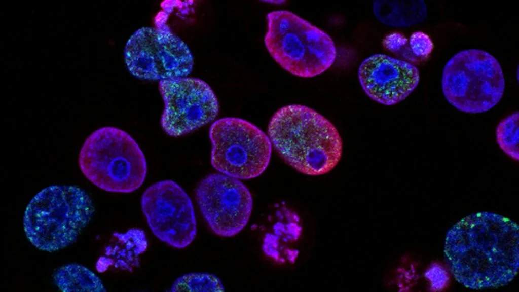
Fluorescence In Situ Hybridization (FISH) is a gold-standard molecular cytogenetic assay utilized to detect chromosomal abnormalities, specifically gene amplification and polysomy, in solid tumors and hematologic malignancies. Quantitative evaluation of fluorescent probe hybridization enables pathologists to establish definitive diagnostic thresholds and select targeted anti-HER2 therapies.
- Primary molecular target: **ERBB2** (HER2) gene locus on chromosome 17q12-q21.
- Diagnostic threshold: **HER2/CEP17 ratio >= 2.0** or absolute gene copy number >= 6.0 signals/cell.
- Therapeutic implication: Direct selection for humanized monoclonal antibodies and antibody-drug conjugates.
Fluorescence in situ hybridization serves as a critical molecular pathology assay for evaluating gene amplification in invasive carcinomas of the breast and gastroesophageal tract. Accurate cytogenetic enumeration directly dictates the deployment of targeted biological therapies in daily clinical oncology practice.
The assay utilizes dual-color fluorescent DNA probes directed against specific target gene loci and centromeric reference chromosomes. Quantitative signal counting identifies true gene amplification versus chromosomal polysomy to guide patient prognosis.
CLINICAL & LABORATORY DIAGNOSTIC PROTOCOL
- Specimen acquisition: Tissue core biopsies or surgical resections fixed immediately in 10% neutral-buffered formalin (NBF) for 6 to 72 hours under strictly controlled cold ischemia times (< 1 hour).
- Slide preparation: 4-micron thick formalin-fixed paraffin-embedded (FFPE) tissue sections mounted on positively charged silanized glass slides baked at 60°C for 60 minutes.
- Control validation: Certified control cell line slides containing known diploid and amplified cells (e.g., MCF-7 and SK-BR-3 breast cancer xenografts) processed concurrently with every batch.
Deparaffinization & Pre-Treatment
Immerse slides in xylene for 3 x 5 minutes, dehydrate through 100% ethanol for 2 x 2 minutes, and air dry. Immerse in 0.2N HCl for 20 minutes, wash in purified water, and boil in pretreatment sodium thiocyanate solution for 30 minutes at 98 degrees Celsius.
Enzymatic Digestion (Proteolysis)
Apply pepsin digestion solution (0.2 mg/mL in 0.01N HCl) pre-warmed to 37 degrees Celsius for 15 to 30 minutes. Rinse thoroughly in 2X SSC (saline-sodium citrate) buffer for 5 minutes at room temperature.
Denaturation & Hybridization
Apply 10 microliters of dual-color DNA probe mixture (**ERBB2 spectrum orange** / **CEP17 spectrum green**) to the targeted tissue area and coverslip with rubber cement sealant. Co-denature the slide and probe on a hotplate at 73 degrees Celsius for 5 minutes, followed by overnight hybridization at 37 degrees Celsius in a humidified chamber.
Post-Hybridization Washes & Nuclear Counterstain
Remove rubber cement and coverslips, wash slides in 0.4X SSC / 0.3% Igepal solution at 72 degrees Celsius for 2 minutes, followed by 2X SSC / 0.1% Igepal for 1 minute at room temperature. Apply 10 microliters of DAPI (4,6-diamidino-2-phenylindole) counterstain to visualize nuclear boundaries.
1. Pathophysiology, Etiology & Cellular Carcinogenesis
Chromosomal instability and focal genomic amplifications represent hallmark mechanisms driving tumorigenesis across multiple epithelial malignancies. Tandem duplication events within specific chromosomal bands lead to massive overexpression of receptor tyrosine kinases and unchecked cellular proliferation.
Epigenetic dysregulation frequently accompanies localized gene amplification, driving aberrant transcription of proto-oncogenes. Histone modifications and localized DNA hypomethylation cooperate to maintain an open chromatin state favorable for sustained transcriptional output.
The tumor microenvironment dynamically influences genomic evolution through selective pressures exerted by hypoxia and metabolic deprivation. Stromal-epithelial crosstalk upregulates angiogenic factors and recruits immunosuppressive myeloid cells to support aggressive clonal expansion.
2. Oncogenic Signal Transduction Pathways & Genomic Alterations
Ligand-independent receptor dimerization triggers constitutive autophosphorylation of intracellular tyrosine residues within the catalytic kinase domain. Structural conformational shifts activate downstream intracellular kinase networks including the **RAS-RAF-MEK-ERK** and **PI3K-AKT-mTOR** pathways.
Aberrant activation of the **PI3K-AKT-mTOR** cascade promotes cellular survival, protein synthesis, and metabolic reprogramming necessary for malignant progression. Negative regulatory loops such as PTEN loss frequently co-occur with **ERBB2** amplification to maximize downstream signal flux.
Secondary mutations within the catalytic kinase domain or downstream effector proteins confer acquired resistance to targeted biological therapies. Upregulation of alternative receptor tyrosine kinases bypasses primary blockade and reactivates downstream survival signaling.
3. Histopathological Grading, Morphological Subtypes & Differential Diagnosis
Invasive carcinomas evaluated by fluorescence in situ hybridization exhibit diverse architectural growth patterns including solid, acinar, papillary, and cribriform arrangements. High-grade nuclear atypia, atypical mitotic figures, and areas of geographic necrosis correlate strongly with underlying gene amplification.
Diagnostic panels utilizing immunohistochemistry and molecular markers such as **TTF-1**, **Napsin A**, and **CK7** distinguish primary adenocarcinomas from squamous cell carcinomas and metastatic lesions. Morphological evaluation ensures that fluorescent scoring is restricted strictly to viable invasive carcinoma cell populations.
Differential diagnosis requires ruling out benign reactive proliferations, glandular dysplasia, and in situ carcinoma components that do not reflect invasive biological potential. Strict adherence to morphologic criteria prevents false-positive scoring of stromal fibroblasts and endothelial cells.
4. Targeted Therapeutics, Landmark Phase III Trials & Resistance Mechanics
Targeted treatment regimens incorporate humanized monoclonal antibodies, tyrosine kinase inhibitors, and antibody-drug conjugates designed to neutralize amplified gene products. Agents such as **trastuzumab**, **pertuzumab**, and **trastuzumab deruxtecan** deliver targeted cytotoxicity directly to cells overexpressing the **ERBB2** receptor.
Landmark Phase III clinical trials including **DESTINY-Breast03** and **CLEOPATRA** established unprecedented improvements in progression-free survival and overall survival for patients harboring confirmed **ERBB2** amplification. Objective response rates remain exceptionally high in biomarker-selected cohorts compared to chemotherapy alone.
Acquired resistance mechanisms involve activating mutations in downstream mediators, steric hindrance of antibody binding sites, and enhanced efflux pump activity. Combination strategies utilizing dual receptor blockade mitigate escape pathways and prolong clinical disease control.
5. Molecular Staging, Minimal Residual Disease (ctDNA) & Prognostic Stratification
Integration of **AJCC 8th edition TNM staging** parameters with quantitative **FISH** biomarker data provides refined risk stratification for patients with localized and metastatic disease. Pathological staging combined with molecular classification optimizes adjuvant and neoadjuvant therapeutic decision-making.
Liquid biopsy surveillance utilizing circulating tumor DNA (**ctDNA**) allows non-invasive tracking of subclonal dynamics and minimal residual disease detection during active therapy. Serial quantification of plasma genomic alterations detects emerging resistance mutations months before radiographic progression.
Prognostic models incorporating quantitative gene copy number ratios and multivariable clinicopathological parameters accurately predict long-term overall survival. Prospective validation of these integrated algorithms continues to refine precision oncology clinical trial design.