Immunohistochemistry (IHC) Staining Protocol for PD-L1 and HER2 Diagnostics in Oncology
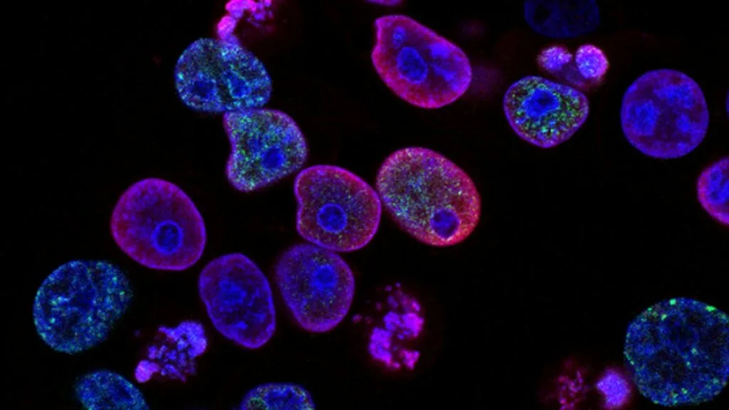
This protocol details the analytical pathology standards for immunohistochemical detection of Programmed Death-Ligand 1 (PD-L1) and Human Epidermal Growth Factor Receptor 2 (HER2). Accurate biomarker quantification directly dictates patient eligibility for monoclonal antibody therapy and antibody-drug conjugates.
- HER2 overexpression driven by ERBB2 amplification requires confirmation via IHC (3+) or ISH (ratio >= 2.0).
- PD-L1 expression quantification requires valid membrane staining on viable tumor cells using standardized clones (22C3, SP263, or 28-8).
- Pre-analytical variables including cold ischemia time strictly impact epitope preservation and diagnostic accuracy.
Immunohistochemical staining standardization is essential for distinguishing true target protein expression from technical artifacts in diagnostic pathology laboratories. Pathologists evaluate distinct cellular compartments, scoring membranous labeling for HER2 and membranous/cytoplasmic labeling for PD-L1.
Targeted oncological management relies heavily on precise companion diagnostics to pair patients with appropriate checkpoint inhibitors or HER2-directed therapies. Analytical failure in pre-analytical tissue handling or post-analytical interpretation directly impairs clinical trial concordance and patient survival.
CLINICAL & LABORATORY DIAGNOSTIC PROTOCOL
- Specimen acquisition: Core needle biopsy or surgical resection fixed in 10% neutral-buffered formalin (NBF) within 30 minutes of devascularization, ensuring cold ischemia time does not exceed 1 hour.
- Tissue processing: Formalin fixation time strictly calibrated to 24-48 hours at room temperature, followed by paraffin embedding and 4-micron microtome sectioning on positively charged slides.
- Control standards: On-slide external positive control tissue with known low/high expression levels and validated negative isotype control run in parallel with every analytical batch.
Tissue Deparaffinization & Heat-Induced Epitope Retrieval (HIER)
Bake slides at 60 degrees Celsius for 60 minutes, deparaffinize through sequential xylene baths (3 changes for 5 minutes each), and rehydrate through graded ethanols (100%, 95%, 70%) to deionized water. Perform HIER in target retrieval solution using pH 9.0 EDTA buffer or pH 6.0 citrate buffer at 97 degrees Celsius for 20 minutes in an automated PT Link module.
Endogenous Peroxidase Block & Primary Antibody Incubation
Quench endogenous peroxidase activity using 3 percent hydrogen peroxide for 10 minutes at room temperature, followed by washing in Tris-buffered saline with Tween-20 (TBST). Apply optimized primary monoclonal antibody clones, such as 22C3 or SP263 for PD-L1 or 4B5 for HER2, at calibrated clinical dilution and incubate for 30 to 60 minutes.
Polymer Detection System & DAB Chromogen Visualization
Rinse slides thoroughly in TBST wash buffer to remove unbound primary antibody. Apply horseradish peroxidase (HRP)-conjugated anti-rabbit or anti-mouse secondary polymer reagent for 30 minutes. Incubate with freshly prepared 3,3-diaminobenzidine (DAB) substrate-chromogen solution for 5 to 10 minutes until brown precipitates develop.
Counterstaining, Dehydration & Quality Control Scoring
Counterstain nuclei with Mayer hematoxylin for 1 to 2 minutes, blue in running tap water, dehydrate through graded ethanols, clear in xylene, and coverslip with permanent non-aqueous mounting medium. Score viable invasive carcinoma cells under light microscopy while excluding necrotic zones and crush artifact.
1. Pathophysiology, Etiology & Cellular Carcinogenesis
Malignant transformation often involves genomic instability and evasion of host immune surveillance mechanisms. Upregulation of inhibitory checkpoint proteins allows neoplastic clones to suppress cytotoxic T-lymphocyte activation within the tumor microenvironment.
Epigenetic dysregulation and oncogene activation drive aberrant cellular proliferation and transmembrane receptor overexpression. Understanding these microenvironmental interactions provides the rationale for combined immunotherapy and targeted cytotoxic delivery.
Stromal composition and infiltrating immune cells heavily influence the spatial distribution of biomarker expression. Pathologists must differentiate between tumor cell membrane staining and intratumoral immune cell staining during evaluation.
2. Oncogenic Signal Transduction Pathways & Genomic Alterations
The **ERBB2** gene encodes a transmembrane receptor tyrosine kinase belonging to the epidermal growth factor receptor family. Amplification of **ERBB2** triggers constitutive homodimerization and ligand-independent activation of downstream mitogenic pathways.
Downstream intracellular signaling proceeds through the **RAS-RAF-MEK-ERK** and **PI3K-AKT-mTOR** cascades, promoting unchecked cellular survival and proliferation. Therapeutic inhibition of these pathways requires accurate diagnostic quantification of receptor density.
Resistance to targeted agents often emerges through secondary mutations, bypass tract activation, or downstream pathway mutations. Molecular pathology assays must identify these alterations to guide subsequent therapeutic lines.
3. Histopathological Grading, Morphological Subtypes & Differential Diagnosis
Histological evaluation requires identifying architectural growth patterns including acinar, papillary, solid, and micropapillary arrangements. Degree of nuclear atypia and mitotic index contribute to overall histological grading and prognostic stratification.
Diagnostic immunohistochemical panels utilizing markers such as **TTF-1**, **Napsin A**, and **CK7** differentiate primary pulmonary adenocarcinomas from metastatic lesions. Negative staining for squamous markers helps confirm lineage specificity.
Accurate morphologic classification prevents diagnostic misallocation between primary tumors and metastatic malignancies. Distinguishing invasive carcinoma from carcinoma in situ ensures only true invasive components are scored for companion diagnostics.
4. Targeted Therapeutics, Landmark Phase III Trials & Resistance Mechanics
Targeted therapies including **trastuzumab deruxtecan**, **pembrolizumab**, and **nivolumab** have revolutionized management algorithms for biomarker-positive malignancies. These agents leverage specific receptor binding and immune restoration to induce tumor regression.
Landmark Phase III clinical investigations, including the **KEYNOTE-024** and **DESTINY-Breast04** trials, demonstrated superior progression-free and overall survival outcomes in biomarker-selected cohorts. These trials established standardized cutoffs for clinical trial inclusion and community practice.
Acquired resistance frequently develops via secondary receptor mutations, loss of surface antigen expression, or upregulation of alternative survival signaling axes. Novel antibody-drug conjugates continue to mitigate resistance by delivering cytotoxic payloads directly to antigen-expressing cells.
5. Molecular Staging, Minimal Residual Disease (ctDNA) & Prognostic Stratification
Integration of **AJCC 8th edition** TNM staging with molecular biomarkers provides a comprehensive framework for patient prognostication. Pathological staging combined with quantitative immunohistochemistry refines recurrence risk assessment.
Liquid biopsy surveillance utilizing circulating tumor DNA (**ctDNA**) enables early detection of minimal residual disease and therapeutic resistance mutations. Serial plasma sampling complements tissue-based immunohistochemical assays.
Overall survival prognostic stratification models incorporate biomarker expression density to predict response duration. Prospective clinical validation ensures that laboratory assays maintain high concordance with patient outcomes.